


Nir2 NirD domain dimer
Rahn, T.A., Lee, W.R., Li, W.T., Airola, M.V., Liou, J.To be published.
Experimental Data Snapshot
Starting Model: experimental
View more details
wwPDB Validation 3D Report Full Report

Entity ID: 1 | |||||
|---|---|---|---|---|---|
| Molecule | Chains | Sequence Length | Organism | Details | Image |
| PITPNM1, Nir2 | 340 | Homo sapiens | Mutation(s): 0 Gene Names: PITPNM1 | 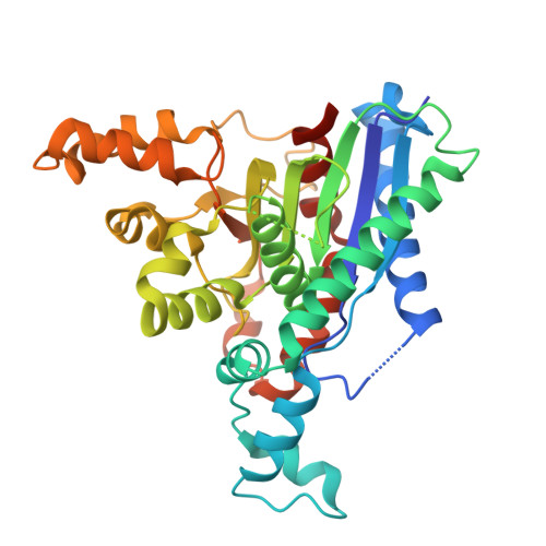 | |
| Length ( Å ) | Angle ( ˚ ) |
|---|---|
| a = 106.765 | α = 90 |
| b = 106.765 | β = 90 |
| c = 136.885 | γ = 120 |
| Software Name | Purpose |
|---|---|
| PHENIX | refinement |
| autoPROC | data reduction |
| STARANISO | data scaling |
| PHENIX | phasing |
| Funding Organization | Location | Grant Number |
|---|---|---|
| National Institutes of Health/National Institute of General Medical Sciences (NIH/NIGMS) | United States | R35GM128666 |


