9C28 | pdb_00009c28

Structure of endogenous Glutamine synthetase from rat model of Alzheimer's disease


Experimental Data Snapshot

  • Method: ELECTRON MICROSCOPY
  • Resolution: 3.12 Å
  • Aggregation State: PARTICLE 
  • Reconstruction Method: SINGLE PARTICLE 

Starting Model: experimental
View more details

wwPDB Validation 3D Report Full Report

Validation slider image for 9C28

This is version 1.1 of the entry. See complete history

Literature

Unveiling the structural proteome of an Alzheimer's disease rat brain model.

Samani, E.K.Hasan, S.M.N.Waas, M.Keszei, A.F.A.Xu, X.Heydari, M.Hill, M.E.McLaurin, J.Kislinger, T.Mazhab-Jafari, M.T.

(2025) Structure 33: 51

  • DOI: https://doi.org/10.1016/j.str.2024.11.004
  • Primary Citation Related Structures: 
    9C21, 9C28, 9C4E, 9C86

  • PubMed Abstract: 

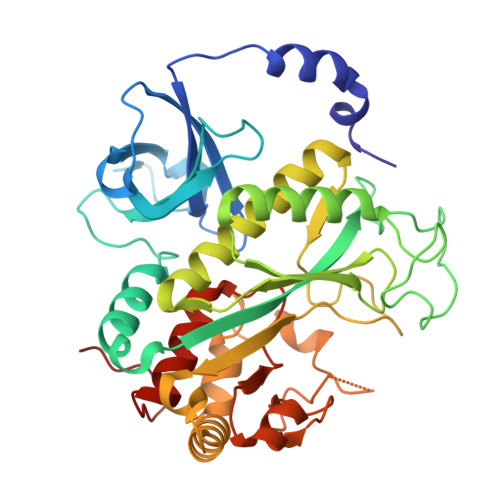
    Studying native protein structures at near-atomic resolution in a crowded environment presents challenges. Consequently, understanding the structural intricacies of proteins within pathologically affected tissues often relies on mass spectrometry and proteomic analysis. Here, we utilized cryoelectron microscopy (cryo-EM) and the Build and Retrieve (BaR) method to investigate protein complexes' structural characteristics such as post-translational modification, active site occupancy, and arrested conformational state in Alzheimer's disease (AD) using brain lysate from a rat model (TgF344-AD). Our findings reveal novel insights into the architecture of these complexes, corroborated through mass spectrometry analysis. Interestingly, it has been shown that the dysfunction of these protein complexes extends beyond AD, implicating them in cancer, as well as other neurodegenerative disorders such as Parkinson's disease, Huntington's disease, and schizophrenia. By elucidating these structural details, our work not only enhances our understanding of disease pathology but also suggests new avenues for future approaches in therapeutic intervention.


  • Organizational Affiliation
    • Department of Medical Biophysics, University of Toronto, Toronto, Ontario, Canada.

Macromolecule Content 

  • Total Structure Weight: 424.34 kDa 
  • Atom Count: 28,010 
  • Modeled Residue Count: 3,680 
  • Deposited Residue Count: 3,730 
  • Unique protein chains: 1

Macromolecules

Find similar proteins by:|  3D Structure
Entity ID: 1
MoleculeChains  Sequence LengthOrganismDetailsImage
Glutamine synthetase
A, B, C, D, E
A, B, C, D, E, F, G, H, I, J
373Rattus norvegicusMutation(s): 0 
EC: 6.3.1.2 (PDB Primary Data), 2.3.1.225 (PDB Primary Data)
UniProt
Find proteins for P09606 (Rattus norvegicus)
Explore P09606 
Go to UniProtKB:  P09606
Entity Groups
Sequence Clusters30% Identity50% Identity70% Identity90% Identity95% Identity100% Identity
UniProt GroupP09606
Sequence Annotations
Expand
Reference Sequence

Small Molecules

Ligands 1 Unique
IDChains Name / Formula / InChI Key2D Diagram3D Interactions
MN
(Subject of Investigation/LOI)

Query on MN


Download:Ideal Coordinates CCD File
AA [auth I]
BA [auth I]
CA [auth J]
DA [auth J]
K [auth A]
AA [auth I],
BA [auth I],
CA [auth J],
DA [auth J],
K [auth A],
L [auth A],
M [auth B],
N [auth B],
O [auth C],
P [auth C],
Q [auth D],
R [auth D],
S [auth E],
T [auth E],
U [auth F],
V [auth F],
W [auth G],
X [auth G],
Y [auth H],
Z [auth H]
MANGANESE (II) ION
Mn
WAEMQWOKJMHJLA-UHFFFAOYSA-N

Experimental Data & Validation

Experimental Data

  • Method: ELECTRON MICROSCOPY
  • Resolution: 3.12 Å
  • Aggregation State: PARTICLE 
  • Reconstruction Method: SINGLE PARTICLE 
EM Software:
TaskSoftware PackageVersion
RECONSTRUCTIONcryoSPARC
MODEL REFINEMENTPHENIX

Structure Validation

View Full Validation Report


Entry History 

& Funding Information

Deposition Data


Funding OrganizationLocationGrant Number
Canadian Institutes of Health Research (CIHR)Canada419240

Revision History  (Full details and data files)

  • Version 1.0: 2024-12-11
    Type: Initial release
  • Version 1.1: 2025-01-15
    Changes: Data collection, Database references