9C13 | pdb_00009c13

XMAP215 TOG5 interaction with GMPCPP tubulin lattice


Experimental Data Snapshot

  • Method: ELECTRON MICROSCOPY
  • Resolution: 2.89 Å
  • Aggregation State: FILAMENT 
  • Reconstruction Method: HELICAL 

Starting Models: in silico, experimental
View more details

wwPDB Validation 3D Report Full Report

Validation slider image for 9C13

This is version 1.1 of the entry. See complete history

Literature

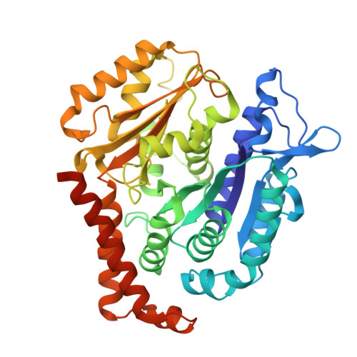
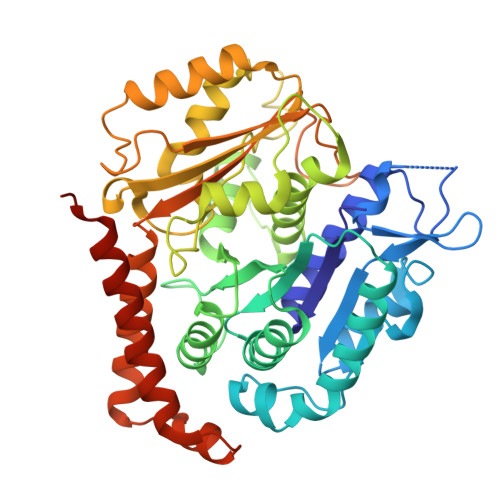
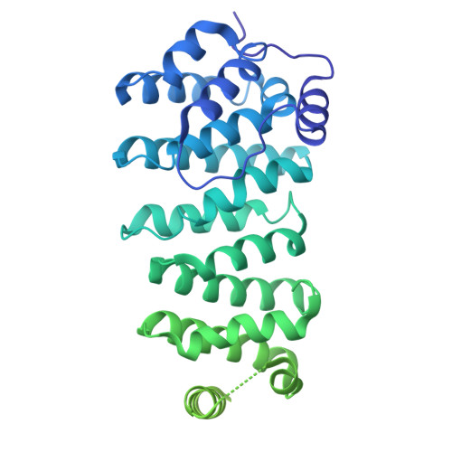
Molecular insight into microtubule nucleation by the XMAP215/ gamma-TuRC module.

McManus, C.T.Travis, S.M.Jeffrey, P.D.Zhang, R.Petry, S.

(2026) Nat Commun 

  • DOI: https://doi.org/10.1038/s41467-026-72370-3
  • Primary Citation Related Structures: 
    9C13

  • PubMed Abstract: 

    It has become increasingly evident in recent years that nucleation of microtubules from a diverse set of microtubule organizing centers (MTOCs) requires both the γ-tubulin ring complex (γ-TuRC) and the microtubule polymerase XMAP215. Despite their essentiality, little is known about how these nucleation factors interact and work together to generate microtubules. Using biochemical domain analysis of XMAP215 and structural approaches, we find that the XMAP215 C-terminal region interacts broadly with γ-TuRC, involving a sixth TOG domain which binds γ-tubulin. Moreover, TOG6 is required for XMAP215 to promote nucleation from γ-TuRC to its full extent. Interestingly, we find that XMAP215 also depends strongly on TOG5 for microtubule lattice binding and nucleation. We further report a cryo-EM structure of TOG5 bound to the microtubule lattice that reveals promotion of lateral interactions between tubulin dimers. While XMAP215 constructs' effects on nucleation are generally proportional to their effects on polymerization, formation of a direct complex with γ-TuRC allows cooperative nucleation activity. Thus, we propose that XMAP215's C-terminal TOGs 5 and 6 play key roles in nucleation by promoting formation of longitudinal and lateral bonds in nascent γ-TuRC-templated microtubules at cellular MTOCs.


  • Organizational Affiliation
    • Department of Molecular Biology, Princeton University, Princeton, NJ, USA.

Macromolecule Content 

  • Total Structure Weight: 264.3 kDa 
  • Atom Count: 15,708 
  • Modeled Residue Count: 1,978 
  • Deposited Residue Count: 2,333 
  • Unique protein chains: 3

Macromolecules

Find similar proteins by:|  3D Structure
Entity ID: 1
MoleculeChains  Sequence LengthOrganismDetailsImage
Tubulin beta-2B chainA [auth B],
B [auth D]
445Bos taurusMutation(s): 0 
UniProt
Find proteins for Q6B856 (Bos taurus)
Explore Q6B856 
Go to UniProtKB:  Q6B856
Entity Groups
Sequence Clusters30% Identity50% Identity70% Identity90% Identity95% Identity100% Identity
UniProt GroupQ6B856
Sequence Annotations
Expand
Reference Sequence
Find similar proteins by:|  3D Structure
Entity ID: 2
MoleculeChains  Sequence LengthOrganismDetailsImage
Tubulin alpha chainC [auth A],
D [auth C]
451Bos taurusMutation(s): 0 
UniProt
Find proteins for F2Z4C1 (Bos taurus)
Explore F2Z4C1 
Go to UniProtKB:  F2Z4C1
Entity Groups
Sequence Clusters30% Identity50% Identity70% Identity90% Identity95% Identity100% Identity
UniProt GroupF2Z4C1
Sequence Annotations
Expand
Reference Sequence
Find similar proteins by:|  3D Structure
Entity ID: 3
MoleculeChains  Sequence LengthOrganismDetailsImage
Cytoskeleton-associated protein 5-A,Cytoskeleton-associated protein 5-A,Green fluorescent protein541Xenopus laevisMutation(s): 0 
Gene Names: ckap5-axmap215GFP
UniProt
Find proteins for Q9PT63 (Xenopus laevis)
Explore Q9PT63 
Go to UniProtKB:  Q9PT63
Entity Groups
Sequence Clusters30% Identity50% Identity70% Identity90% Identity95% Identity100% Identity
UniProt GroupQ9PT63
Sequence Annotations
Expand
Reference Sequence

Small Molecules

Ligands 3 Unique
IDChains Name / Formula / InChI Key2D Diagram3D Interactions
GTP

Query on GTP


Download:Ideal Coordinates CCD File
J [auth A],
L [auth C]
GUANOSINE-5'-TRIPHOSPHATE
C10 H16 N5 O14 P3
XKMLYUALXHKNFT-UUOKFMHZSA-N
G2P

Query on G2P


Download:Ideal Coordinates CCD File
F [auth B],
H [auth D]
PHOSPHOMETHYLPHOSPHONIC ACID GUANYLATE ESTER
C11 H18 N5 O13 P3
GXTIEXDFEKYVGY-KQYNXXCUSA-N
MG

Query on MG


Download:Ideal Coordinates CCD File
G [auth B],
I [auth D],
K [auth A],
M [auth C]
MAGNESIUM ION
Mg
JLVVSXFLKOJNIY-UHFFFAOYSA-N

Experimental Data & Validation

Experimental Data

  • Method: ELECTRON MICROSCOPY
  • Resolution: 2.89 Å
  • Aggregation State: FILAMENT 
  • Reconstruction Method: HELICAL 
EM Software:
TaskSoftware PackageVersion
MODEL REFINEMENTPHENIX1.21-5207-000
RECONSTRUCTIONcryoSPARC3.3.2

Structure Validation

View Full Validation Report


Entry History 

& Funding Information

Deposition Data


Funding OrganizationLocationGrant Number
National Institutes of Health/National Institute of General Medical Sciences (NIH/NIGMS)United States1R01 GM141100-01A1
National Institutes of Health/National Institute of General Medical Sciences (NIH/NIGMS)United StatesF32 GM142149-01A1
National Institutes of Health/National Institute of General Medical Sciences (NIH/NIGMS)United States1R01 GM141100-01A1

Revision History  (Full details and data files)

  • Version 1.0: 2025-11-05
    Type: Initial release
  • Version 1.1: 2026-05-20
    Changes: Data collection, Database references