9BVY | pdb_00009bvy

Neutron Structure of Peroxide-Soaked Tyr34Phe MnSOD


Experimental Data Snapshot

  • Method: NEUTRON DIFFRACTION
  • Resolution: 2.30 Å
  • R-Value Free: 
    0.299 (Depositor) 
  • R-Value Work: 
    0.265 (Depositor) 
  • R-Value Observed: 
    0.267 (Depositor) 

Starting Model: experimental
View more details

wwPDB Validation 3D Report Full Report

Validation slider image for 9BVY

This is version 1.0 of the entry. See complete history

Literature

The role of Tyr34 in proton coupled electron transfer and product inhibition of manganese superoxide dismutase.

Azadmanesh, J.Slobodnik, K.Struble, L.R.Lovelace, J.J.Cone, E.A.Dasgupta, M.Lutz, W.E.Kumar, S.Natarajan, A.Coates, L.Weiss, K.L.Myles, D.A.A.Kroll, T.Borgstahl, G.E.O.

(2025) Nat Commun 16: 1887-1887

  • DOI: https://doi.org/10.1038/s41467-025-57180-3
  • Primary Citation Related Structures: 
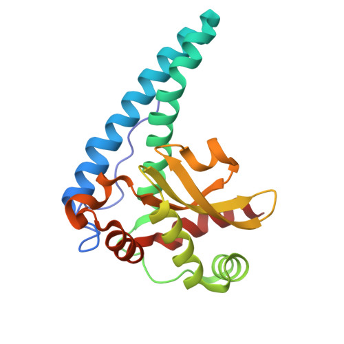
    9BVY, 9BW2, 9BWM, 9BWQ, 9BWR

  • PubMed Abstract: 

    Human manganese superoxide dismutase (MnSOD) plays a crucial role in controlling levels of reactive oxygen species (ROS) by converting superoxide ( O 2 - ) to molecular oxygen (O 2 ) and hydrogen peroxide (H 2 O 2 ) with proton-coupled electron transfers (PCETs). A key catalytic residue, Tyr34, determines the activity of human MnSOD and also becomes post-translationally inactivated by nitration in various diseases associated with mitochondrial dysfunction. Tyr34 has an unusual pK a due to its proximity to the Mn metal and undergoes cyclic deprotonation and protonation events to promote the electron transfers of MnSOD. Neutron diffraction, X-ray spectroscopy, and quantum chemistry calculations in oxidized, reduced and product inhibited enzymatic states shed light on the role of Tyr34 in MnSOD catalysis. The data identify the contributions of Tyr34 in MnSOD activity that support mitochondrial function and give a thorough characterization of how a single tyrosine modulates PCET catalysis. Product inhibition occurs by an associative displacement mechanism.


  • Organizational Affiliation
    • Eppley Institute for Research in Cancer and Allied Diseases, 986805 Nebraska Medical Center, Omaha, NE, USA.

Macromolecule Content 

  • Total Structure Weight: 44.84 kDa 
  • Atom Count: 3,664 
  • Modeled Residue Count: 398 
  • Deposited Residue Count: 398 
  • Unique protein chains: 1

Macromolecules

Find similar proteins by:|  3D Structure
Entity ID: 1
MoleculeChains  Sequence LengthOrganismDetailsImage
Superoxide dismutase [Mn], mitochondrial
A, B
199Homo sapiensMutation(s): 1 
Gene Names: SOD2
EC: 1.15.1.1
UniProt & NIH Common Fund Data Resources
Find proteins for P04179 (Homo sapiens)
Explore P04179 
Go to UniProtKB:  P04179
PHAROS:  P04179
Entity Groups
Sequence Clusters30% Identity50% Identity70% Identity90% Identity95% Identity100% Identity
UniProt GroupP04179
Sequence Annotations
Expand
Reference Sequence

Experimental Data & Validation

Experimental Data

  • Method: NEUTRON DIFFRACTION
  • Resolution: 2.30 Å
  • R-Value Free:  0.299 (Depositor) 
  • R-Value Work:  0.265 (Depositor) 
  • R-Value Observed: 0.267 (Depositor) 
Space Group: P 61 2 2
Unit Cell:
Length ( Å )Angle ( ˚ )
a = 78.69α = 90
b = 78.69β = 90
c = 239.94γ = 120

Structure Validation

View Full Validation Report


Entry History 

& Funding Information

Deposition Data


Funding OrganizationLocationGrant Number
National Institutes of Health/National Institute of General Medical Sciences (NIH/NIGMS)United StatesR01-GM145647
National Aeronautics and Space Administration (NASA, United States)United States44-0307-1021-201
Department of Energy (DOE, United States)United States--

Revision History  (Full details and data files)

  • Version 1.0: 2025-03-12
    Type: Initial release