9BUK

Outward-facing, ATP-bound Multidrug Resistance-associated Protein 2 (MRP2) (E1462Q)


Experimental Data Snapshot

  • Method: ELECTRON MICROSCOPY
  • Resolution: 3.40 Å
  • Aggregation State: PARTICLE 
  • Reconstruction Method: SINGLE PARTICLE 

wwPDB Validation   3D Report Full Report


This is version 1.1 of the entry. See complete history


Literature

Structural basis for the transport and regulation mechanism of the multidrug resistance-associated protein 2.

Koide, E.Pietz, H.L.Beltran, J.Chen, J.

(2025) Nat Commun 16: 484-484

  • DOI: https://doi.org/10.1038/s41467-024-55810-w
  • Primary Citation of Related Structures:  
    9BR2, 9BUK, 9C12, 9C2I

  • PubMed Abstract: 

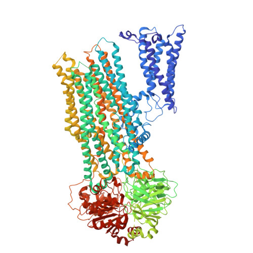
    Multidrug resistance-associated protein 2 (MRP2) is an ATP-powered exporter important for maintaining liver homeostasis and a potential contributor to chemotherapeutic resistance. Using cryogenic electron microscopy (cryo-EM), we determine the structures of human MRP2 in three conformational states: an autoinhibited state, a substrate-bound pre-translocation state, and an ATP-bound post-translocation state. In the autoinhibited state, the cytosolic regulatory (R) domain plugs into the transmembrane substrate-binding site and extends into the cytosol to form a composite ATP-binding site at the surface of nucleotide-binding domain 2. Substrate displaces the R domain, permitting conformational changes necessary for transport. These observations suggest that the R domain functions as a selectivity gauge, where only at sufficiently high concentrations can the substrate effectively initiate transport. Comparative structural analyzes of MRP2 bound to various substrates, as determined in this study and others, reveal how MRP2 recognizes a diverse array of compounds, supporting its role in multidrug resistance.


  • Organizational Affiliation

    Laboratory of Membrane Biology and Biophysics, The Rockefeller University, New York, NY, USA.


Macromolecules
Find similar proteins by:  (by identity cutoff)  |  3D Structure
Entity ID: 1
MoleculeChains Sequence LengthOrganismDetailsImage
ATP-binding cassette sub-family C member 21,545Homo sapiensMutation(s): 1 
Gene Names: ABCC2CMOATCMOAT1CMRPMRP2
EC: 7.6.2 (PDB Primary Data), 7.6.2.2 (PDB Primary Data), 7.6.2.3 (PDB Primary Data)
Membrane Entity: Yes 
UniProt & NIH Common Fund Data Resources
Find proteins for Q92887 (Homo sapiens)
Explore Q92887 
Go to UniProtKB:  Q92887
PHAROS:  Q92887
GTEx:  ENSG00000023839 
Entity Groups  
Sequence Clusters30% Identity50% Identity70% Identity90% Identity95% Identity100% Identity
UniProt GroupQ92887
Sequence Annotations
Expand
  • Reference Sequence
Small Molecules
Ligands 4 Unique
IDChains Name / Formula / InChI Key2D Diagram3D Interactions
ATP (Subject of Investigation/LOI)
Query on ATP

Download Ideal Coordinates CCD File 
B [auth A],
C [auth A]
ADENOSINE-5'-TRIPHOSPHATE
C10 H16 N5 O13 P3
ZKHQWZAMYRWXGA-KQYNXXCUSA-N
CLR
Query on CLR

Download Ideal Coordinates CCD File 
I [auth A],
J [auth A],
K [auth A],
L [auth A],
M [auth A]
CHOLESTEROL
C27 H46 O
HVYWMOMLDIMFJA-DPAQBDIFSA-N
MG
Query on MG

Download Ideal Coordinates CCD File 
D [auth A],
E [auth A]
MAGNESIUM ION
Mg
JLVVSXFLKOJNIY-UHFFFAOYSA-N
UNL
Query on UNL

Download Ideal Coordinates CCD File 
F [auth A],
G [auth A],
H [auth A]
Unknown ligand
JLVVSXFLKOJNIY-UHFFFAOYSA-N
Experimental Data & Validation

Experimental Data

  • Method: ELECTRON MICROSCOPY
  • Resolution: 3.40 Å
  • Aggregation State: PARTICLE 
  • Reconstruction Method: SINGLE PARTICLE 
EM Software:
TaskSoftware PackageVersion
MODEL REFINEMENTPHENIX1.21.1_5286

Structure Validation

View Full Validation Report


Entry History & Funding Information

Deposition Data


Funding OrganizationLocationGrant Number
Howard Hughes Medical Institute (HHMI)United States--

Revision History  (Full details and data files)

  • Version 1.0: 2024-12-18
    Type: Initial release
  • Version 1.1: 2025-02-12
    Changes: Data collection, Database references