9BTS

Crystal structure of the bacterioferritin (Bfr) and ferritin (Ftn) heterooligomer complex from Acinetobacter baumannii


Experimental Data Snapshot

  • Method: X-RAY DIFFRACTION
  • Resolution: 1.85 Å
  • R-Value Free: 0.199 
  • R-Value Work: 0.166 
  • R-Value Observed: 0.168 

Starting Model: experimental
View more details

wwPDB Validation   3D Report Full Report


Ligand Structure Quality Assessment 


This is version 1.1 of the entry. See complete history


Literature

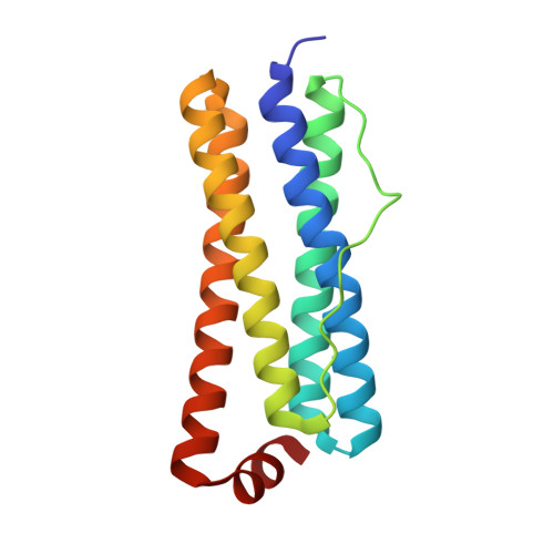
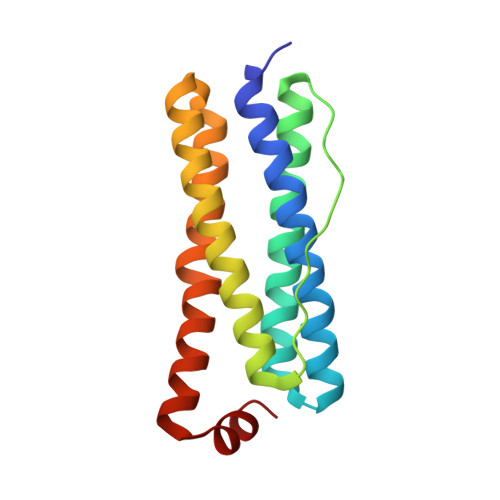
The crystal structure of Acinetobacter baumannii bacterioferritin reveals a heteropolymer of bacterioferritin and ferritin subunits.

Yao, H.Alli, S.Liu, L.Soldano, A.Cooper, A.Fontenot, L.Verdin, D.Battaile, K.P.Lovell, S.Rivera, M.

(2024) Sci Rep 14: 18242-18242

  • DOI: https://doi.org/10.1038/s41598-024-69156-2
  • Primary Citation of Related Structures:  
    9BTS

  • PubMed Abstract: 

    Iron storage proteins, e.g., vertebrate ferritin, and the ferritin-like bacterioferritin (Bfr) and bacterial ferritin (Ftn), are spherical, hollow proteins that catalyze the oxidation of Fe 2+ at binuclear iron ferroxidase centers (FOC) and store the Fe 3+ in their interior, thus protecting cells from unwanted Fe 3+ /Fe 2+ redox cycling and storing iron at concentrations far above the solubility of Fe 3+ . Vertebrate ferritins are heteropolymers of H and L subunits with only the H subunits having FOC. Bfr and Ftn were thought to coexist in bacteria as homopolymers, but recent evidence indicates these molecules are heteropolymers assembled from Bfr and Ftn subunits. Despite the heteropolymeric nature of vertebrate and bacterial ferritins, structures have been determined only for recombinant proteins constituted by a single subunit type. Herein we report the structure of Acinetobacter baumannii bacterioferritin, the first structural example of a heteropolymeric ferritin or ferritin-like molecule, assembled from completely overlapping Ftn homodimers harboring FOC and Bfr homodimers devoid of FOC but binding heme. The Ftn homodimers function by catalyzing the oxidation of Fe 2+ to Fe 3+ , while the Bfr homodimers bind a cognate ferredoxin (Bfd) which reduces the stored Fe 3+ by transferring electrons via the heme, enabling Fe 2+ mobilization to the cytosol for incorporation in metabolism.


  • Organizational Affiliation

    Department of Chemistry, Louisiana State University, Baton Rouge, 70803, USA.


Macromolecules
Find similar proteins by:  (by identity cutoff)  |  3D Structure
Entity ID: 1
MoleculeChains Sequence LengthOrganismDetailsImage
Bacterioferritin (Bfr)
A, B, C, D, E
A, B, C, D, E, F, G, H
154Acinetobacter baumannii ATCC 17978Mutation(s): 0 
EC: 1.16.3.1
UniProt
Find proteins for A0A1E3M9R5 (Acinetobacter baumannii)
Explore A0A1E3M9R5 
Go to UniProtKB:  A0A1E3M9R5
Entity Groups  
Sequence Clusters30% Identity50% Identity70% Identity90% Identity95% Identity100% Identity
UniProt GroupA0A1E3M9R5
Sequence Annotations
Expand
  • Reference Sequence
Find similar proteins by:  (by identity cutoff)  |  3D Structure
Entity ID: 2
MoleculeChains Sequence LengthOrganismDetailsImage
Ferritin (Ftn)154Acinetobacter baumannii ATCC 17978Mutation(s): 0 
EC: 1.16.3.1
UniProt
Find proteins for T2HRY3 (Acinetobacter baumannii)
Explore T2HRY3 
Go to UniProtKB:  T2HRY3
Entity Groups  
Sequence Clusters30% Identity50% Identity70% Identity90% Identity95% Identity100% Identity
UniProt GroupT2HRY3
Sequence Annotations
Expand
  • Reference Sequence
Small Molecules
Ligands 2 Unique
IDChains Name / Formula / InChI Key2D Diagram3D Interactions
HEM (Subject of Investigation/LOI)
Query on HEM

Download Ideal Coordinates CCD File 
S [auth A],
T [auth C],
W [auth F],
Y [auth H]
PROTOPORPHYRIN IX CONTAINING FE
C34 H32 Fe N4 O4
KABFMIBPWCXCRK-RGGAHWMASA-L
SO4
Query on SO4

Download Ideal Coordinates CCD File 
AA [auth c]
BA [auth e]
CA [auth g]
DA [auth g]
Q [auth A]
AA [auth c],
BA [auth e],
CA [auth g],
DA [auth g],
Q [auth A],
R [auth A],
U [auth D],
V [auth F],
X [auth H],
Z [auth b]
SULFATE ION
O4 S
QAOWNCQODCNURD-UHFFFAOYSA-L
Experimental Data & Validation

Experimental Data

  • Method: X-RAY DIFFRACTION
  • Resolution: 1.85 Å
  • R-Value Free: 0.199 
  • R-Value Work: 0.166 
  • R-Value Observed: 0.168 
  • Space Group: P 2 3
Unit Cell:
Length ( Å )Angle ( ˚ )
a = 173.028α = 90
b = 173.028β = 90
c = 173.028γ = 90
Software Package:
Software NamePurpose
PHENIXrefinement
Aimlessdata scaling
XDSdata reduction
PHASERphasing

Structure Validation

View Full Validation Report


Ligand Structure Quality Assessment 


Entry History & Funding Information

Deposition Data


Funding OrganizationLocationGrant Number
National Institutes of Health/Office of the DirectorUnited StatesS10OD030394
National Institutes of Health/National Institute Of Allergy and Infectious Diseases (NIH/NIAID)United StatesAI169344

Revision History  (Full details and data files)

  • Version 1.0: 2024-08-14
    Type: Initial release
  • Version 1.1: 2024-08-21
    Changes: Database references