9BEQ | pdb_00009beq

Crystal structure of pregnane X receptor ligand binding domain complexed with AP1867


Experimental Data Snapshot

  • Method: X-RAY DIFFRACTION
  • Resolution: 2.60 Å
  • R-Value Free: 
    0.246 (Depositor), 0.245 (DCC) 
  • R-Value Work: 
    0.215 (Depositor), 0.215 (DCC) 
  • R-Value Observed: 
    0.217 (Depositor) 

Starting Model: experimental
View more details

wwPDB Validation 3D Report Full Report

Validation slider image for 9BEQ

Ligand Structure Quality Assessment 


This is version 1.2 of the entry. See complete history

Literature

PROTAC-mediated activation, rather than degradation, of a nuclear receptor reveals complex ligand-receptor interaction network.

Huber, A.D.Lin, W.Poudel, S.Miller, D.J.Chen, T.

(2024) Structure 32: 2352-2363.e8

  • DOI: https://doi.org/10.1016/j.str.2024.09.016
  • Primary Citation Related Structures: 
    9BEQ

  • PubMed Abstract: 

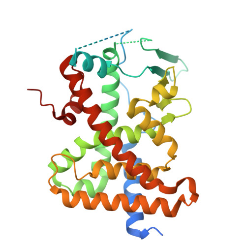
    Proteolysis-targeting chimeras (PROTACs) are heterobifunctional molecules containing a ligand for a protein of interest linked to an E3 ubiquitin ligase ligand that induce protein degradation through E3 recruitment to the target protein. Small changes in PROTAC linkers can have drastic consequences, including loss of degradation activity, but the structural mechanisms governing such changes are unclear. To study this phenomenon, we screened PROTACs of diverse targeting modalities and identified dTAG-13 as an activator of the xenobiotic-sensing pregnane X receptor (PXR), which promiscuously binds various ligands. Characterization of dTAG-13 analogs and precursors revealed interplay between the PXR-binding moiety, linker, and E3 ligand that altered PXR activity without inducing degradation. A crystal structure of PXR ligand binding domain bound to a precursor ligand showed ligand-induced binding pocket distortions and a linker-punctured tunnel to the protein exterior at a region incompatible with E3 complex formation, highlighting the effects of linker environment on PROTAC activity.


  • Organizational Affiliation
    • Department of Chemical Biology and Therapeutics, St. Jude Children's Research Hospital, 262 Danny Thomas Place, MS 1000, Memphis, TN 38105-3678, USA.

Macromolecule Content 

  • Total Structure Weight: 36.9 kDa 
  • Atom Count: 2,191 
  • Modeled Residue Count: 267 
  • Deposited Residue Count: 316 
  • Unique protein chains: 1

Macromolecules

Find similar proteins by:|  3D Structure
Entity ID: 1
MoleculeChains  Sequence LengthOrganismDetailsImage
Nuclear receptor subfamily 1 group I member 2316Homo sapiensMutation(s): 0 
Gene Names: NR1I2PXR
UniProt & NIH Common Fund Data Resources
Find proteins for O75469 (Homo sapiens)
Explore O75469 
Go to UniProtKB:  O75469
PHAROS:  O75469
GTEx:  ENSG00000144852 
Entity Groups
Sequence Clusters30% Identity50% Identity70% Identity90% Identity95% Identity100% Identity
UniProt GroupO75469
Sequence Annotations
Expand
Reference Sequence

Small Molecules

Ligands 1 Unique
IDChains Name / Formula / InChI Key2D Diagram3D Interactions
AP1
(Subject of Investigation/LOI)

Query on AP1


Download:Ideal Coordinates CCD File
B [auth A]{3-[3-(3,4-DIMETHOXY-PHENYL)-1-(1-{1-[2-(3,4,5-TRIMETHOXY-PHENYL)-BUTYRYL]-PIPERIDIN-2YL}-VINYLOXY)-PROPYL]-PHENOXY}-ACETIC ACID
C38 H47 N O11
XCCRAOPQCACRFC-OIFRRMEBSA-N

Experimental Data & Validation

Experimental Data

  • Method: X-RAY DIFFRACTION
  • Resolution: 2.60 Å
  • R-Value Free:  0.246 (Depositor), 0.245 (DCC) 
  • R-Value Work:  0.215 (Depositor), 0.215 (DCC) 
  • R-Value Observed: 0.217 (Depositor) 
Space Group: P 43 21 2
Unit Cell:
Length ( Å )Angle ( ˚ )
a = 91.33α = 90
b = 91.33β = 90
c = 85.26γ = 90
Software Package:
Software NamePurpose
PHENIXrefinement
XDSdata reduction
XDSdata scaling
PHASERphasing
PDB_EXTRACTdata extraction

Structure Validation

View Full Validation Report


Ligand Structure Quality Assessment 


Entry History 

& Funding Information

Deposition Data


Funding OrganizationLocationGrant Number
National Institutes of Health/National Institute of General Medical Sciences (NIH/NIGMS)United StatesR35GM118041

Revision History  (Full details and data files)

  • Version 1.0: 2024-10-09
    Type: Initial release
  • Version 1.1: 2024-10-23
    Changes: Database references
  • Version 1.2: 2024-12-18
    Changes: Database references