9B8G | pdb_00009b8g

2C9 Fab antibody fragment against the E protein of the Yellow Fever virus


Experimental Data Snapshot

  • Method: X-RAY DIFFRACTION
  • Resolution: 2.66 Å
  • R-Value Free: 
    0.258 (Depositor), 0.258 (DCC) 
  • R-Value Work: 
    0.201 (Depositor), 0.200 (DCC) 
  • R-Value Observed: 
    0.203 (Depositor) 

Starting Model: experimental
View more details

wwPDB Validation 3D Report Full Report

Validation slider image for 9B8G

This is version 1.1 of the entry. See complete history

Literature

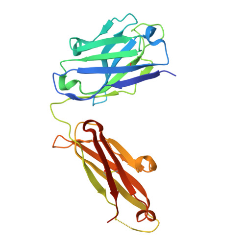
A single residue in the yellow fever virus envelope protein modulates virion architecture and antigenicity.

Bibby, S.Jung, J.Low, Y.S.Amarilla, A.A.Newton, N.D.Scott, C.A.P.Balk, J.Ting, Y.T.Freney, M.E.Liang, B.Grant, T.Coulibaly, F.Young, P.Hall, R.A.Hobson-Peters, J.Modhiran, N.Watterson, D.

(2025) Nat Commun 16: 8449-8449

  • DOI: https://doi.org/10.1038/s41467-025-63038-5
  • Primary Citation Related Structures: 
    9B6U, 9B6V, 9B6W, 9B6X, 9B6Y, 9B8G

  • PubMed Abstract: 

    Yellow fever virus (YFV) is a re-emerging flavivirus that causes severe hepatic disease and mortality in humans. Despite being researched for over a century, the structure of YFV has remained elusive. Here we use a chimeric virus platform to resolve the first high resolution cryo-EM structures of YFV. Stark differences in particle morphology and homogeneity are observed between vaccine and virulent strains of YFV, and these are found to have significant implications on antibody recognition and neutralisation. We identify a single residue (R380) in the YFV 17D envelope protein that stabilises the virion surface, and leads to reduced exposure of the cross-reactive fusion loop epitope. The differences in virion morphology between YFV strains also contribute to the reduced sensitivity of the virulent YFV virions to vaccine-induced antibodies. These findings have significant implications for YFV biology, vaccinology and structure-based flavivirus antigen design.


  • Organizational Affiliation
    • School of Chemistry & Molecular Biosciences, The University of Queensland, St Lucia, QLD, Australia.

Macromolecule Content 

  • Total Structure Weight: 146.85 kDa 
  • Atom Count: 10,200 
  • Modeled Residue Count: 1,296 
  • Deposited Residue Count: 1,344 
  • Unique protein chains: 2

Macromolecules

Find similar proteins by:|  3D Structure
Entity ID: 1
MoleculeChains  Sequence LengthOrganismDetailsImage
2C9 Fab heavy chainA,
C,
E [auth H]
230Homo sapiensMutation(s): 0 
Find similar proteins by:|  3D Structure
Entity ID: 2
MoleculeChains  Sequence LengthOrganismDetailsImage
2C9 Fab light chainB,
D,
F [auth L]
218Homo sapiensMutation(s): 0 

Experimental Data & Validation

Experimental Data

  • Method: X-RAY DIFFRACTION
  • Resolution: 2.66 Å
  • R-Value Free:  0.258 (Depositor), 0.258 (DCC) 
  • R-Value Work:  0.201 (Depositor), 0.200 (DCC) 
  • R-Value Observed: 0.203 (Depositor) 
Space Group: P 1 21 1
Unit Cell:
Length ( Å )Angle ( ˚ )
a = 67.667α = 90
b = 125.329β = 93.947
c = 96.928γ = 90
Software Package:
Software NamePurpose
PHENIXrefinement
XDSdata reduction
Aimlessdata scaling
PHASERphasing

Structure Validation

View Full Validation Report


Entry History 

& Funding Information

Deposition Data


Funding OrganizationLocationGrant Number
National Health and Medical Research Council (NHMRC, Australia)AustraliaAPP1164216
National Health and Medical Research Council (NHMRC, Australia)AustraliaAPP2004582

Revision History  (Full details and data files)

  • Version 1.0: 2024-11-06
    Type: Initial release
  • Version 1.1: 2025-10-15
    Changes: Database references