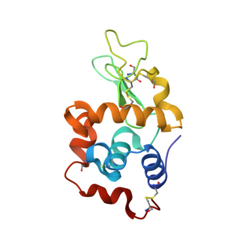
9B7C | pdb_00009b7c

high-resolution ambient temperature structure of lysozyme soaked with sodium iodide

  • Classification: HYDROLASE
  • Organism(s): Gallus gallus
  • Mutation(s): No 

  • Deposited: 2024-03-27 Released: 2024-07-31 
  • Deposition Author(s): Wang, H.K., Greisman, J.B., Hekstra, D.R.
  • Funding Organization(s): National Science Foundation (NSF, United States), National Institutes of Health/Office of the Director

Experimental Data Snapshot

  • Method: X-RAY DIFFRACTION
  • Resolution: 1.10 Å
  • R-Value Free: 
    0.123 (Depositor), 0.122 (DCC) 
  • R-Value Work: 
    0.111 (Depositor), 0.110 (DCC) 
  • R-Value Observed: 
    0.111 (Depositor) 

wwPDB Validation 3D Report Full Report

Validation slider image for 9B7C

This is version 1.2 of the entry. See complete history

Literature

Sensitive detection of structural dynamics using a statistical framework for comparative crystallography.

Hekstra, D.R.Wang, H.K.Klureza, M.A.Greisman, J.B.Dalton, K.M.

(2025) Sci Adv 11: eadj2921-eadj2921

  • DOI: https://doi.org/10.1126/sciadv.adj2921
  • Primary Citation Related Structures: 
    9B7C

  • PubMed Abstract: 

    Chemical and conformational changes are crucial to protein function and its pharmacological control. X-ray crystallography can reveal these changes in atomic detail, but standard analysis methods, which refine separate datasets, often overlook differences that are subtle or arise in only a subset of molecules. Direct comparison of crystallographic datasets is, in principle, more powerful, but systematic errors ("scales") often mask changes in the crystallographic observables ("structure factors"). Machine learning algorithms that jointly estimate scales and structure factors can address this limitation. Here, we augment this approach with multivariate, structured priors derived from crystallographic theory, implemented in the variational deep learning framework Careless. Doing so strongly improves the detection of protein dynamics, element-specific anomalous signals, and the binding of drug candidates, offering a robust approach to comparative crystallography and, potentially, to detection of protein dynamics by other structure determination methods.


  • Organizational Affiliation
    • Department of Molecular and Cellular Biology, Harvard University, Cambridge, MA 02138, USA.

Macromolecule Content 

  • Total Structure Weight: 14.92 kDa 
  • Atom Count: 1,284 
  • Modeled Residue Count: 129 
  • Deposited Residue Count: 129 
  • Unique protein chains: 1

Macromolecules

Find similar proteins by:|  3D Structure
Entity ID: 1
MoleculeChains  Sequence LengthOrganismDetailsImage
Lysozyme C129Gallus gallusMutation(s): 0 
EC: 3.2.1.17
UniProt
Find proteins for P00698 (Gallus gallus)
Explore P00698 
Go to UniProtKB:  P00698
Entity Groups
Sequence Clusters30% Identity50% Identity70% Identity90% Identity95% Identity100% Identity
UniProt GroupP00698
Sequence Annotations
Expand
Reference Sequence

Experimental Data & Validation

Experimental Data

  • Method: X-RAY DIFFRACTION
  • Resolution: 1.10 Å
  • R-Value Free:  0.123 (Depositor), 0.122 (DCC) 
  • R-Value Work:  0.111 (Depositor), 0.110 (DCC) 
  • R-Value Observed: 0.111 (Depositor) 
Space Group: P 43 21 2
Unit Cell:
Length ( Å )Angle ( ˚ )
a = 79.405α = 90
b = 79.405β = 90
c = 37.837γ = 90
Software Package:
Software NamePurpose
PHENIXrefinement
Aimlessdata scaling
DIALSdata reduction
AutoSolphasing

Structure Validation

View Full Validation Report


Entry History 

& Funding Information

Deposition Data


Funding OrganizationLocationGrant Number
National Science Foundation (NSF, United States)United StatesDGE 2140743
National Science Foundation (NSF, United States)United StatesDGE1745303
National Institutes of Health/Office of the DirectorUnited StatesDP2-GM141000

Revision History  (Full details and data files)

  • Version 1.0: 2024-07-31
    Type: Initial release
  • Version 1.1: 2024-10-30
    Changes: Structure summary
  • Version 1.2: 2026-02-11
    Changes: Database references