9B2D | pdb_00009b2d

Yeast Rad51-ssDNA filament


Experimental Data Snapshot

  • Method: ELECTRON MICROSCOPY
  • Resolution: 2.40 Å
  • Aggregation State: FILAMENT 
  • Reconstruction Method: SINGLE PARTICLE 

Starting Model: experimental
View more details

wwPDB Validation 3D Report Full Report

Validation slider image for 9B2D

This is version 1.1 of the entry. See complete history

Literature

Local structural dynamics of Rad51 protomers revealed by cryo-electron microscopy of Rad51-ssDNA filaments.

Liu, J.Gore, S.K.Heyer, W.D.

(2025) Nucleic Acids Res 53

  • DOI: https://doi.org/10.1093/nar/gkaf052
  • Primary Citation Related Structures: 
    9B2D, 9ED3

  • PubMed Abstract: 

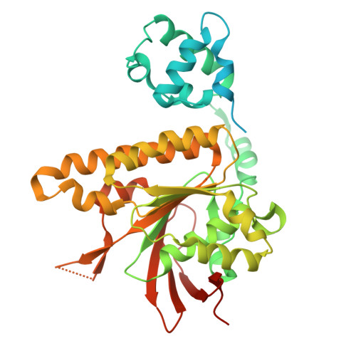
    Homologous recombination (HR) is a high-fidelity repair mechanism for double-strand breaks. Rad51 is the key enzyme that forms filaments on single-stranded DNA (ssDNA) to catalyze homology search and DNA strand exchange in recombinational DNA repair. In this study, we employed single-particle cryogenic electron microscopy (cryo-EM) to ascertain the density map of the wild-type budding yeast Rad51-ssDNA filament bound to ADP-AlF3, achieving a resolution of 2.35 Å without imposing helical symmetry. The model assigned 6 Rad51 protomers, 24 nt of DNA, and 6 bound ADP-AlF3. It shows 6-fold symmetry implying monomeric building blocks, unlike the structure of the Rad51-I345T mutant filament with three-fold symmetry implying dimeric building blocks, for which the structural comparisons provide a satisfying mechanistic explanation. This image analysis enables comprehensive comparisons of individual Rad51 protomers within the filament and reveals local conformational movements of amino acid side chains. Notably, R293 in Loop 1 adopts multiple conformations to facilitate L296 and V331 in separating and twisting the DNA triplets. We also analyzed the crystal structure of Rad51-I345T and the predicted structure of yeast Rad51-K342E using the Rad51-ssDNA structure from this study as a reference.


  • Organizational Affiliation
    • Department of Microbiology & Molecular Genetics, University of California, Davis, Davis, CA 95616-8665, United States.

Macromolecule Content 

  • Total Structure Weight: 268.51 kDa 
  • Atom Count: 14,766 
  • Modeled Residue Count: 1,878 
  • Deposited Residue Count: 2,424 
  • Unique protein chains: 1
  • Unique nucleic acid chains: 1

Macromolecules


Find similar proteins by:|  3D Structure
Entity ID: 1
MoleculeChains  Sequence LengthOrganismDetailsImage
DNA repair protein RAD51400Saccharomyces cerevisiae S288CMutation(s): 0 
Gene Names: RAD51YER095W
UniProt
Find proteins for P25454 (Saccharomyces cerevisiae (strain ATCC 204508 / S288c))
Explore P25454 
Go to UniProtKB:  P25454
Entity Groups
Sequence Clusters30% Identity50% Identity70% Identity90% Identity95% Identity100% Identity
UniProt GroupP25454
Sequence Annotations
Expand
Reference Sequence
Find similar nucleic acids by:  Sequence
Entity ID: 2
MoleculeChains LengthOrganismImage
DNA (5'-D(P*TP*TP*TP*TP*TP*TP*TP*TP*TP*TP*TP*TP*TP*TP*TP*TP*TP*TP*TP*TP*TP*TP*TP*T)-3')G [auth K]24Saccharomyces cerevisiae
Sequence Annotations
Expand
Reference Sequence

Small Molecules

Ligands 3 Unique
IDChains Name / Formula / InChI Key2D Diagram3D Interactions
ADP
(Subject of Investigation/LOI)

Query on ADP


Download:Ideal Coordinates CCD File
H [auth C]
K [auth D]
N [auth E]
Q [auth F]
T [auth A]
H [auth C],
K [auth D],
N [auth E],
Q [auth F],
T [auth A],
W [auth B]
ADENOSINE-5'-DIPHOSPHATE
C10 H15 N5 O10 P2
XTWYTFMLZFPYCI-KQYNXXCUSA-N
AF3
(Subject of Investigation/LOI)

Query on AF3


Download:Ideal Coordinates CCD File
I [auth C]
L [auth D]
O [auth E]
R [auth F]
U [auth A]
I [auth C],
L [auth D],
O [auth E],
R [auth F],
U [auth A],
X [auth B]
ALUMINUM FLUORIDE
Al F3
KLZUFWVZNOTSEM-UHFFFAOYSA-K
MG
(Subject of Investigation/LOI)

Query on MG


Download:Ideal Coordinates CCD File
J [auth C]
M [auth D]
P [auth E]
S [auth F]
V [auth A]
J [auth C],
M [auth D],
P [auth E],
S [auth F],
V [auth A],
Y [auth B]
MAGNESIUM ION
Mg
JLVVSXFLKOJNIY-UHFFFAOYSA-N

Experimental Data & Validation

Experimental Data

  • Method: ELECTRON MICROSCOPY
  • Resolution: 2.40 Å
  • Aggregation State: FILAMENT 
  • Reconstruction Method: SINGLE PARTICLE 
EM Software:
TaskSoftware PackageVersion
RECONSTRUCTIONcryoSPARC3.2
MODEL REFINEMENTPHENIX1.20.1

Structure Validation

View Full Validation Report


Entry History 

& Funding Information

Deposition Data


Funding OrganizationLocationGrant Number
National Institutes of Health/National Institute of General Medical Sciences (NIH/NIGMS)United StatesGM58015
National Institutes of Health/National Institute of General Medical Sciences (NIH/NIGMS)United StatesGM137751
National Institutes of Health/National Cancer Institute (NIH/NCI)United StatesP30CA093373
National Institutes of Health/National Institute of General Medical Sciences (NIH/NIGMS)United StatesU24 GM129541

Revision History  (Full details and data files)

  • Version 1.0: 2025-01-29
    Type: Initial release
  • Version 1.1: 2026-02-11
    Changes: Data collection, Database references