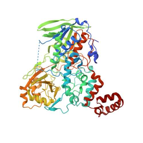
Cryo-EM reconstruction of oleate hydratase bound to a phospholipid membrane bilayer.
Oldham, M.L., Zuhaib Qayyum, M., Kalathur, R.C., Rock, C.O., Radka, C.D.(2024) J Struct Biol 216: 108116-108116
- PubMed: 39151742 Search on PubMed
- DOI: https://doi.org/10.1016/j.jsb.2024.108116
- Primary Citation Related Structures:
8UR8, 9AXE - PubMed Abstract:
Oleate hydratase (OhyA) is a bacterial peripheral membrane protein that catalyzes FAD-dependent water addition to membrane bilayer-embedded unsaturated fatty acids. The opportunistic pathogen Staphylococcus aureus uses OhyA to counteract the innate immune system and support colonization. Many Gram-positive and Gram-negative bacteria in the microbiome also encode OhyA. OhyA is a dimeric flavoenzyme whose carboxy terminus is identified as the membrane binding domain; however, understanding how OhyA binds to cellular membranes is not complete until the membrane-bound structure has been elucidated. All available OhyA structures depict the solution state of the protein outside its functional environment. Here, we employ liposomes to solve the cryo-electron microscopy structure of the functional unit: the OhyA•membrane complex. The protein maintains its structure upon membrane binding and slightly alters the curvature of the liposome surface. OhyA preferentially associates with 20-30 nm liposomes with multiple copies of OhyA dimers assembling on the liposome surface resulting in the formation of higher-order oligomers. Dimer assembly is cooperative and extends along a formed ridge of the liposome. We also solved an OhyA dimer of dimers structure that recapitulates the intermolecular interactions that stabilize the dimer assembly on the membrane bilayer as well as the crystal contacts in the lattice of the OhyA crystal structure. Our work enables visualization of the molecular trajectory of membrane binding for this important interfacial enzyme.
- Department of Structural Biology, St. Jude Children's Research Hospital, Memphis, TN, 38105, USA.
Organizational Affiliation: