8ZPU | pdb_00008zpu

Crystal structure of the anti-phosphorylated peptide C7 scFv antibody with peptide bound


Experimental Data Snapshot

  • Method: X-RAY DIFFRACTION
  • Resolution: 2.80 Å
  • R-Value Free: 
    0.345 (Depositor), 0.347 (DCC) 
  • R-Value Work: 
    0.304 (Depositor), 0.307 (DCC) 

Starting Model: experimental
View more details

wwPDB Validation   3D Report Full Report


This is version 1.1 of the entry. See complete history


Literature

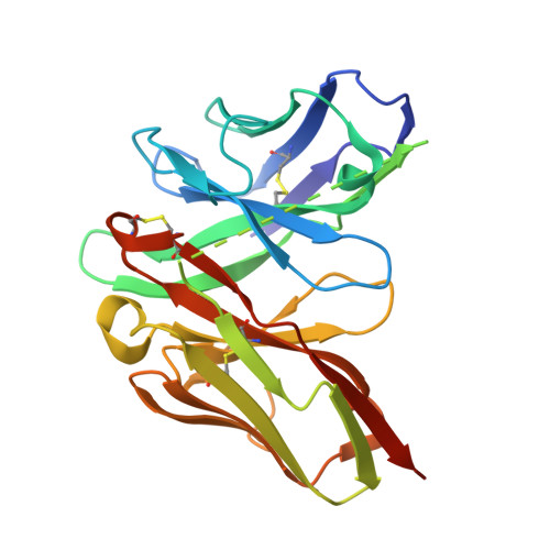
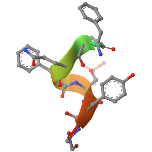
Unveiling the structural mechanisms behind high affinity and selectivity in phosphorylated epitope-specific rabbit antibodies.

Kasahara, K.Kawade, R.Nakakido, M.Matsunaga, R.Akiba, H.Entzminger, K.C.Maruyama, T.Okumura, S.C.J.Caaveiro, J.M.M.Kuroda, D.Tsumoto, K.

(2024) J Biological Chem 300: 107989-107989

  • DOI: https://doi.org/10.1016/j.jbc.2024.107989
  • Primary Citation of Related Structures:  
    8JOW, 8ZPU, 8ZXW

  • PubMed Abstract: 

    Protein phosphorylation is a crucial process in various cellular functions, and its irregularities have been implicated in several diseases, including cancer. Antibodies are commonly employed to detect protein phosphorylation in research. However, unlike the extensive studies on recognition mechanisms of the phosphate group by proteins such as kinases and phosphatases, only a few studies have explored antibody mechanisms. In this study, we produced and characterized two rabbit monoclonal antibodies that recognize a monophosphorylated Akt peptide. Through crystallography, thermodynamic mutational analyses, and molecular dynamics simulations, we investigated the unique recognition mechanism that enables higher binding affinity and selectivity of the antibodies compared to other generic proteins with lower binding affinity to phosphorylated epitopes. Our results demonstrate that molecular dynamics simulations provide novel insights into the dynamic aspects of molecular recognition of posttranslational modifications by proteins beyond static crystal structures, highlighting how specific atomic level interactions drive the exceptional affinity and selectivity of antibodies.


  • Organizational Affiliation

    Department of Bioengineering, School of Engineering, The University of Tokyo, Bunkyo-ku, Tokyo, Japan.


Macromolecules
Find similar proteins by:  (by identity cutoff)  |  3D Structure
Entity ID: 1
MoleculeChains Sequence LengthOrganismDetailsImage
scFvA,
C [auth B]
257Oryctolagus cuniculusMutation(s): 0 
Entity Groups  
Sequence Clusters30% Identity50% Identity70% Identity90% Identity95% Identity100% Identity
Sequence Annotations
Expand
  • Reference Sequence

Find similar proteins by:  Sequence   |   3D Structure  

Entity ID: 2
MoleculeChains Sequence LengthOrganismDetailsImage
PHE-PRO-GLN-PHE-SEP-TYR-SERB [auth C],
D
12Homo sapiensMutation(s): 0 
EC: 2.7.11.1
UniProt & NIH Common Fund Data Resources
Find proteins for P31749 (Homo sapiens)
Explore P31749 
Go to UniProtKB:  P31749
PHAROS:  P31749
GTEx:  ENSG00000142208 
Entity Groups  
Sequence Clusters30% Identity50% Identity70% Identity90% Identity95% Identity100% Identity
UniProt GroupP31749
Sequence Annotations
Expand
  • Reference Sequence
Small Molecules
Ligands 2 Unique
IDChains Name / Formula / InChI Key2D Diagram3D Interactions
SO4
Query on SO4

Download Ideal Coordinates CCD File 
E [auth A]
F [auth A]
G [auth A]
I [auth B]
J [auth B]
SULFATE ION
O4 S
QAOWNCQODCNURD-UHFFFAOYSA-L
CL
Query on CL

Download Ideal Coordinates CCD File 
H [auth A],
L [auth B]
CHLORIDE ION
Cl
VEXZGXHMUGYJMC-UHFFFAOYSA-M
Modified Residues  1 Unique
IDChains TypeFormula2D DiagramParent
SEP
Query on SEP
B [auth C],
D
L-PEPTIDE LINKINGC3 H8 N O6 PSER
Experimental Data & Validation

Experimental Data

  • Method: X-RAY DIFFRACTION
  • Resolution: 2.80 Å
  • R-Value Free:  0.345 (Depositor), 0.347 (DCC) 
  • R-Value Work:  0.304 (Depositor), 0.307 (DCC) 
Space Group: P 32 2 1
Unit Cell:
Length ( Å )Angle ( ˚ )
a = 126.61α = 90
b = 126.61β = 90
c = 82.36γ = 120
Software Package:
Software NamePurpose
REFMACrefinement
MOSFLMdata reduction
Aimlessdata scaling
PHASERphasing

Structure Validation

View Full Validation Report


Entry History & Funding Information

Deposition Data


Funding OrganizationLocationGrant Number
Japan Society for the Promotion of Science (JSPS)Japan15K06962

Revision History  (Full details and data files)

  • Version 1.0: 2024-12-11
    Type: Initial release
  • Version 1.1: 2024-12-25
    Changes: Database references