8YYR

Structure of the HitB T293G mutant


Experimental Data Snapshot

  • Method: X-RAY DIFFRACTION
  • Resolution: 1.80 Å
  • R-Value Free: 0.211 
  • R-Value Work: 0.177 
  • R-Value Observed: 0.178 

wwPDB Validation   3D Report Full Report


Ligand Structure Quality Assessment 


This is version 1.0 of the entry. See complete history


Literature

Engineering the Substrate Specificity of (S)-beta-Phenylalanine Adenylation Enzyme HitB.

Wang, D.Miyanaga, A.Chisuga, T.Kudo, F.Eguchi, T.

(2024) Chembiochem : e202400383-e202400383

  • DOI: https://doi.org/10.1002/cbic.202400383
  • Primary Citation of Related Structures:  
    8YYQ, 8YYR

  • PubMed Abstract: 

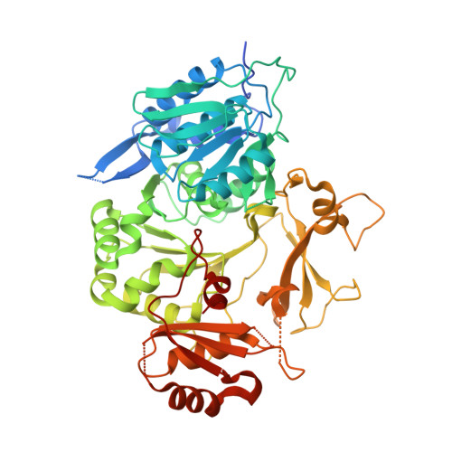
    Adenylation enzymes catalyze the selective incorporation of aminoacyl building blocks in the biosynthesis of nonribosomal peptides and related natural products. Although β-amino acid units are one of the important aminoacyl building blocks in natural product biosynthesis, very little is known about the engineering of β-amino acid adenylation enzymes. In this study, we engineered the substrate specificity of the (S)-β-phenylalanine adenylation enzyme, HitB, involved in the biosynthesis of macrolactam polyketide hitachimycin. Based on the previously determined structure of HitB wild-type, we mutated Phe328 and Ser293, which are located near the meta and ortho position of the (S)-β-phenylalanine moiety, respectively. As a result, the HitB F328V and F328L mutants efficiently activated meta-substituted (S)-β-phenylalanine analogs, and the HitB T293G and T293S mutants efficiently activated ortho-substituted (S)-β-phenylalanine analogs. Structural analysis of the HitB F328L and T293G mutants with the corresponding nonhydrolyzable intermediate analogs revealed an enlarged substrate binding pocket for (S)-β-phenylalanine analogs, providing detailed insights into the structural basis for creating enzyme substrate promiscuity. Our findings may be useful for production of various β-amino acid-containing natural product analogs.


  • Organizational Affiliation

    Tokyo Institute of Technology, Department of Chemistry, JAPAN.


Macromolecules
Find similar proteins by:  (by identity cutoff)  |  3D Structure
Entity ID: 1
MoleculeChains Sequence LengthOrganismDetailsImage
Putative ATP-dependent b-aminoacyl-ACP synthetase549Embleya scabrisporaMutation(s): 1 
Gene Names: hitB
UniProt
Find proteins for A0A0F7R6G7 (Embleya scabrispora)
Explore A0A0F7R6G7 
Go to UniProtKB:  A0A0F7R6G7
Entity Groups  
Sequence Clusters30% Identity50% Identity70% Identity90% Identity95% Identity100% Identity
UniProt GroupA0A0F7R6G7
Sequence Annotations
Expand
  • Reference Sequence
Small Molecules
Ligands 1 Unique
IDChains Name / Formula / InChI Key2D Diagram3D Interactions
A1L0H (Subject of Investigation/LOI)
Query on A1L0H

Download Ideal Coordinates CCD File 
B [auth A][(2~{R},3~{S},4~{R},5~{R})-5-(6-aminopurin-9-yl)-3,4-bis(oxidanyl)oxolan-2-yl]methyl ~{N}-[(3~{S})-3-azanyl-3-(2-bromophenyl)propanoyl]sulfamate
C19 H22 Br N7 O7 S
GOEQSWCMWTVHGV-NSDPQSHHSA-N
Experimental Data & Validation

Experimental Data

  • Method: X-RAY DIFFRACTION
  • Resolution: 1.80 Å
  • R-Value Free: 0.211 
  • R-Value Work: 0.177 
  • R-Value Observed: 0.178 
  • Space Group: P 31 2 1
Unit Cell:
Length ( Å )Angle ( ˚ )
a = 67.88α = 90
b = 67.88β = 90
c = 196.654γ = 120
Software Package:
Software NamePurpose
REFMACrefinement
PDB_EXTRACTdata extraction
XDSdata reduction
Aimlessdata scaling
MOLREPphasing

Structure Validation

View Full Validation Report


Ligand Structure Quality Assessment 


Entry History & Funding Information

Deposition Data


Funding OrganizationLocationGrant Number
Not funded--

Revision History  (Full details and data files)

  • Version 1.0: 2024-06-05
    Type: Initial release