8YRG | pdb_00008yrg

CryoEM structure of fospropofol-bound MRGPRX4-Gq complex


Experimental Data Snapshot

  • Method: ELECTRON MICROSCOPY
  • Resolution: 3.14 Å
  • Aggregation State: PARTICLE 
  • Reconstruction Method: SINGLE PARTICLE 

wwPDB Validation 3D Report Full Report

Validation slider image for 8YRG

This is version 1.2 of the entry. See complete history

Literature

MRGPRX4 mediates phospho-drug-associated pruritus in a humanized mouse model.

Chien, D.C.Limjunyawong, N.Cao, C.Meixiong, J.Peng, Q.Ho, C.Y.Fay, J.F.Roth, B.L.Dong, X.

(2024) Sci Transl Med 16: eadk8198-eadk8198

  • DOI: https://doi.org/10.1126/scitranslmed.adk8198
  • Primary Citation Related Structures: 
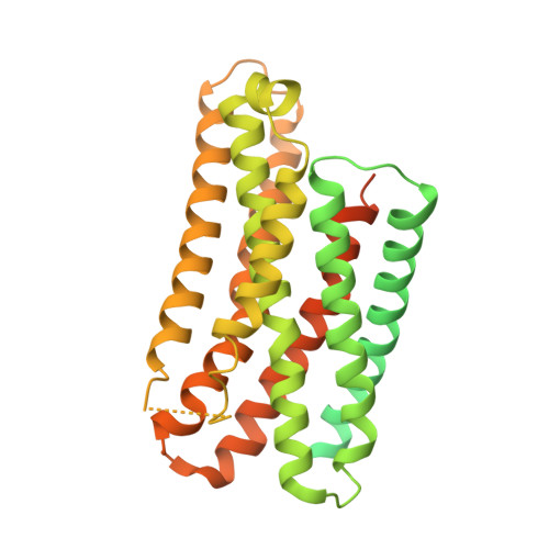
    8YRG

  • PubMed Abstract: 

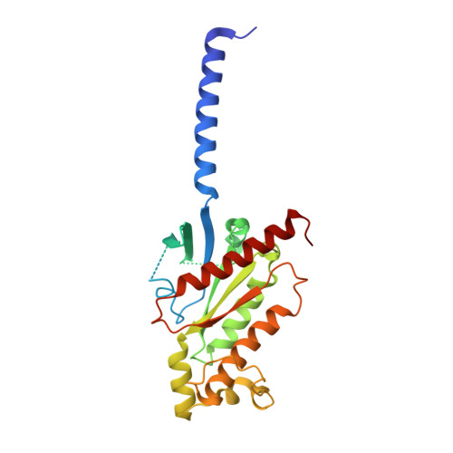
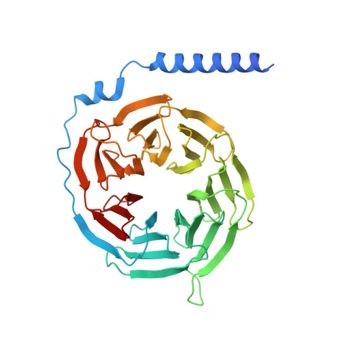
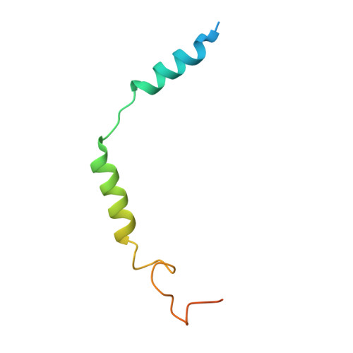
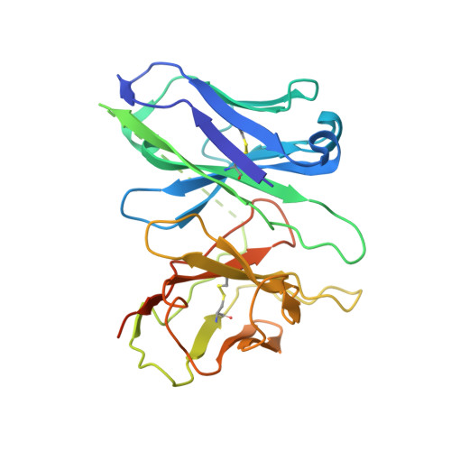
    The phosphate modification of drugs is a common chemical strategy to increase solubility and allow for parenteral administration. Unfortunately, phosphate modifications often elicit treatment- or dose-limiting pruritus through an unknown mechanism. Using unbiased high-throughput drug screens, we identified the Mas-related G protein-coupled receptor X4 (MRGPRX4), a primate-specific, sensory neuron receptor previously implicated in itch, as a potential target for phosphate-modified compounds. Using both G q -mediated calcium mobilization and G protein-independent GPCR assays, we found that phosphate-modified compounds potently activate MRGPRX4. Furthermore, a humanized mouse model expressing MRGPRX4 in sensory neurons exhibited robust phosphomonoester prodrug-evoked itch. To characterize and confirm this interaction, we further determined the structure of MRGPRX4 in complex with a phosphate-modified drug through single-particle cryo-electron microscopy (cryo-EM) and identified critical amino acid residues responsible for the binding of the phosphate group. Together, these findings explain how phosphorylated drugs can elicit treatment-limiting itch and identify MRGPRX4 as a potential therapeutic target to suppress itch and to guide future drug design.


  • Organizational Affiliation
    • Solomon H. Snyder Department of Neuroscience, Johns Hopkins University School of Medicine, Baltimore, MD 21205, USA.

Macromolecule Content 

  • Total Structure Weight: 157.72 kDa 
  • Atom Count: 7,977 
  • Modeled Residue Count: 1,079 
  • Deposited Residue Count: 1,414 
  • Unique protein chains: 5

Macromolecules

Find similar proteins by:|  3D Structure
Entity ID: 1
MoleculeChains  Sequence LengthOrganismDetailsImage
Gs-mini-Gq chimeraA [auth B]246Homo sapiensMutation(s): 0 
Find similar proteins by:|  3D Structure
Entity ID: 2
MoleculeChains  Sequence LengthOrganismDetailsImage
Guanine nucleotide-binding protein G(I)/G(S)/G(T) subunit beta-1B [auth C]358Homo sapiensMutation(s): 0 
Gene Names: GNB1
UniProt & NIH Common Fund Data Resources
Find proteins for P62873 (Homo sapiens)
Explore P62873 
Go to UniProtKB:  P62873
PHAROS:  P62873
GTEx:  ENSG00000078369 
Entity Groups
Sequence Clusters30% Identity50% Identity70% Identity90% Identity95% Identity100% Identity
UniProt GroupP62873
Sequence Annotations
Expand
Reference Sequence
Find similar proteins by:|  3D Structure
Entity ID: 3
MoleculeChains  Sequence LengthOrganismDetailsImage
Guanine nucleotide-binding protein G(I)/G(S)/G(O) subunit gamma-2C [auth D]71Homo sapiensMutation(s): 0 
Gene Names: GNG2
UniProt & NIH Common Fund Data Resources
Find proteins for P59768 (Homo sapiens)
Explore P59768 
Go to UniProtKB:  P59768
PHAROS:  P59768
GTEx:  ENSG00000186469 
Entity Groups
Sequence Clusters30% Identity50% Identity70% Identity90% Identity95% Identity100% Identity
UniProt GroupP59768
Sequence Annotations
Expand
Reference Sequence
Find similar proteins by:|  3D Structure
Entity ID: 4
MoleculeChains  Sequence LengthOrganismDetailsImage
scFv16D [auth E]267Mus musculusMutation(s): 0 
Find similar proteins by:|  3D Structure
Entity ID: 5
MoleculeChains  Sequence LengthOrganismDetailsImage
Soluble cytochrome b562,Mas-related G-protein coupled receptor member X4E [auth R]472Homo sapiensMutation(s): 1 
Gene Names: cybCMRGPRX4MRGX4SNSR5SNSR6
UniProt & NIH Common Fund Data Resources
Find proteins for P0ABE7 (Escherichia coli)
Explore P0ABE7 
Go to UniProtKB:  P0ABE7
Find proteins for Q96LA9 (Homo sapiens)
Explore Q96LA9 
Go to UniProtKB:  Q96LA9
PHAROS:  Q96LA9
GTEx:  ENSG00000179817 
Entity Groups
Sequence Clusters30% Identity50% Identity70% Identity90% Identity95% Identity100% Identity
UniProt GroupsQ96LA9P0ABE7
Sequence Annotations
Expand
Reference Sequence

Small Molecules

Ligands 1 Unique
IDChains Name / Formula / InChI Key2D Diagram3D Interactions
A1LZU
(Subject of Investigation/LOI)

Query on A1LZU


Download:Ideal Coordinates CCD File
F [auth R][2,6-di(propan-2-yl)phenoxy]methyl dihydrogen phosphate
C13 H21 O5 P
QVNNONOFASOXQV-UHFFFAOYSA-N

Experimental Data & Validation

Experimental Data

  • Method: ELECTRON MICROSCOPY
  • Resolution: 3.14 Å
  • Aggregation State: PARTICLE 
  • Reconstruction Method: SINGLE PARTICLE 
EM Software:
TaskSoftware PackageVersion
MODEL REFINEMENTPHENIX

Structure Validation

View Full Validation Report


Entry History 

& Funding Information

Deposition Data


Funding OrganizationLocationGrant Number
National Institutes of Health/National Institute of Diabetes and Digestive and Kidney Disease (NIH/NIDDK)United StatesR01DA055656
National Institutes of Health/National Institute of Diabetes and Digestive and Kidney Disease (NIH/NIDDK)United StatesU24DK116195

Revision History  (Full details and data files)

  • Version 1.0: 2024-05-22
    Type: Initial release
  • Version 1.1: 2024-10-16
    Changes: Data collection, Structure summary
  • Version 1.2: 2025-06-18
    Changes: Data collection