Mechanistic Analysis of Riboswitch Ligand Interactions Provides Insights into Pharmacological Control over Gene Expression.
Parmar, S., Bume, D.D., Conelly, C., Boer, R., Prestwood, P.R., Wang, Z., Labuhn, H., Sinnadurai, K., Feri, A., Ouellet, J., Homan, P., Numata, T., Schneekloth Jr., J.S.(2024) bioRxiv
- PubMed: 38903087 Search on PubMedSearch on PubMed Central
- DOI: https://doi.org/10.1101/2024.02.23.581746
- Primary Citation Related Structures:
8YAM, 8YAN - PubMed Abstract:
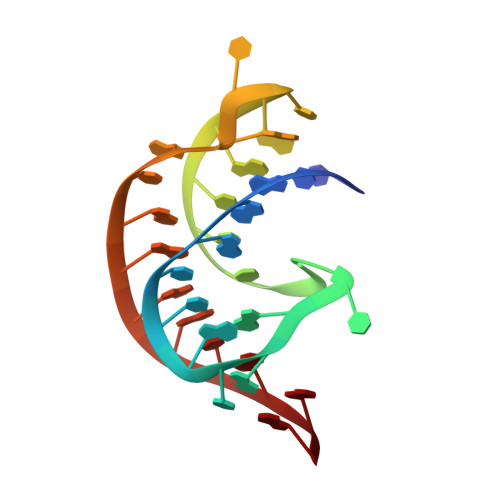
Riboswitches are structured RNA elements that regulate gene expression upon binding to small molecule ligands. Understanding the mechanisms by which small molecules impact riboswitch activity is key to developing potent, selective ligands for these and other RNA targets. We report the structure-informed design of chemically diverse synthetic ligands for PreQ 1 riboswitches. Multiple X-ray co-crystal structures of synthetic ligands with the Thermoanaerobacter tengcongensis ( Tte )-PreQ 1 riboswitch confirm a common binding site with the cognate ligand, despite considerable chemical differences among the ligands. Structure probing assays demonstrate that one ligand causes conformational changes similar to PreQ 1 in six structurally and mechanistically diverse PreQ 1 riboswitch aptamers. Single-molecule force spectroscopy is used to demonstrate differential modes of riboswitch stabilization by the ligands. Binding of the natural ligand brings about the formation of a persistent, folded pseudoknot structure, whereas a synthetic ligand decreases the rate of unfolding through a kinetic mechanism. Single round transcription termination assays show the biochemical activity of the ligands, while a GFP reporter system reveals compound activity in regulating gene expression in live cells without toxicity. Taken together, this study reveals that diverse small molecules can impact gene expression in live cells by altering conformational changes in RNA structures through distinct mechanisms.
- Chemical Biology Laboratory, Center for Cancer Research, National Cancer Institute, Frederick, MD 21702-1201, USA.
Organizational Affiliation: