8XIA | pdb_00008xia

X-RAY ANALYSIS OF D-XYLOSE ISOMERASE AT 1.9 ANGSTROMS: NATIVE ENZYME IN COMPLEX WITH SUBSTRATE AND WITH A MECHANISM-DESIGNED INACTIVATOR


Experimental Data Snapshot

  • Method: X-RAY DIFFRACTION
  • Resolution: 1.90 Å
  • R-Value Observed: 
    0.140 (Depositor) 

wwPDB Validation 3D Report Full Report

Validation slider image for 8XIA

This is version 1.4 of the entry. See complete history

Literature

X-ray analysis of D-xylose isomerase at 1.9 A: native enzyme in complex with substrate and with a mechanism-designed inactivator.

Carrell, H.L.Glusker, J.P.Burger, V.Manfre, F.Tritsch, D.Biellmann, J.F.

(1989) Proc Natl Acad Sci U S A 86: 4440-4444

  • DOI: https://doi.org/10.1073/pnas.86.12.4440
  • Primary Citation Related Structures: 
    8XIA, 9XIA

  • PubMed Abstract: 

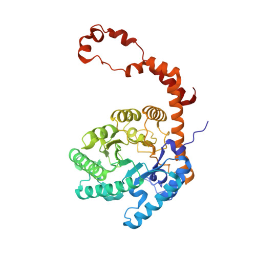
    The structures of crystalline D-xylose isomerase (D-xylose ketol-isomerase; EC 5.3.1.5) from Streptomyces rubiginosus and of its complexes with substrate and with an active-site-directed inhibitor have been determined by x-ray diffraction techniques and refined to 1.9-A resolution. This study identifies the active site, as well as two metal-binding sites. The metal ions are important in maintaining the structure of the active-site region and one of them binds C3-O and C5-O of the substrate forming a six-membered ring. This study has revealed a very close contact between histidine and C1 of a substrate, suggesting that this is the active-site base that abstracts a proton from substrate. The mechanism-based inhibitor is a substrate analog and is turned over by the enzyme to give a product that alkylates this same histidine, reinforcing our interpretation. The changes in structure of the native enzyme, the enzyme with bound substrate, and the alkylated enzyme indicate that the mechanism involves an "open-chain" conformation of substrate and that the intermediate in the isomerization reaction is probably a cis-ene diol because the active-site histidine is correctly placed to abstract a proton from C1 or C2 of the substrate. A water molecule binds to C1O and C2O of the substrate and so may act as a proton donor or acceptor in the enolization of a ring-opened substrate.


  • Organizational Affiliation
    • Institute for Cancer Research, Fox Chase Cancer Center, Philadelphia, PA 19111.

Macromolecule Content 

  • Total Structure Weight: 43.51 kDa 
  • Atom Count: 3,347 
  • Modeled Residue Count: 387 
  • Deposited Residue Count: 388 
  • Unique protein chains: 1

Macromolecules

Find similar proteins by:|  3D Structure
Entity ID: 1
MoleculeChains  Sequence LengthOrganismDetailsImage
XYLOSE ISOMERASE388Streptomyces pseudogriseolusMutation(s): 0 
EC: 5.3.1.5
UniProt
Find proteins for P24300 (Streptomyces pseudogriseolus)
Explore P24300 
Go to UniProtKB:  P24300
Entity Groups
Sequence Clusters30% Identity50% Identity70% Identity90% Identity95% Identity100% Identity
UniProt GroupP24300
Sequence Annotations
Expand
Reference Sequence

Experimental Data & Validation

Experimental Data

  • Method: X-RAY DIFFRACTION
  • Resolution: 1.90 Å
  • R-Value Observed: 0.140 (Depositor) 
Space Group: I 2 2 2
Unit Cell:
Length ( Å )Angle ( ˚ )
a = 94.2α = 90
b = 100.2β = 90
c = 103.2γ = 90
Software Package:
Software NamePurpose
PROLSQrefinement

Structure Validation

View Full Validation Report


Entry History 

Deposition Data

Revision History  (Full details and data files)

  • Version 1.0: 1991-10-15
    Type: Initial release
  • Version 1.1: 2008-03-25
    Changes: Version format compliance
  • Version 1.2: 2011-07-13
    Changes: Derived calculations, Version format compliance
  • Version 1.3: 2020-07-29
    Type: Remediation
    Reason: Carbohydrate remediation
    Changes: Database references, Derived calculations, Other, Structure summary
  • Version 1.4: 2024-02-14
    Changes: Data collection, Database references, Structure summary