8XBH

Human GPR34 -Gi complex bound to M1


Experimental Data Snapshot

  • Method: ELECTRON MICROSCOPY
  • Resolution: 2.83 Å
  • Aggregation State: PARTICLE 
  • Reconstruction Method: SINGLE PARTICLE 

wwPDB Validation   3D Report Full Report


This is version 1.1 of the entry. See complete history


Literature

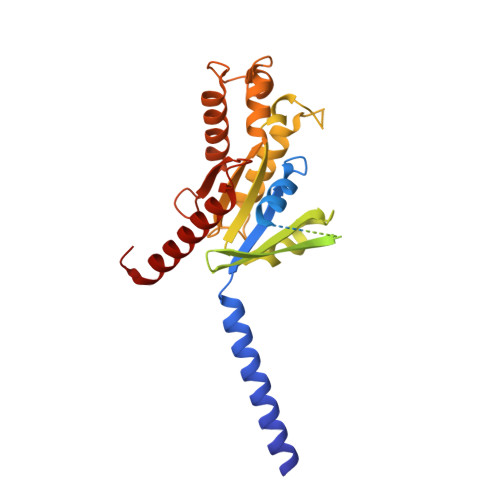
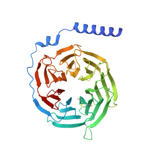
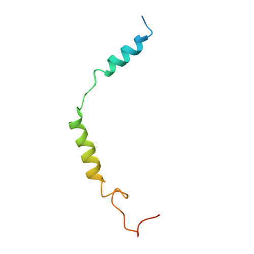
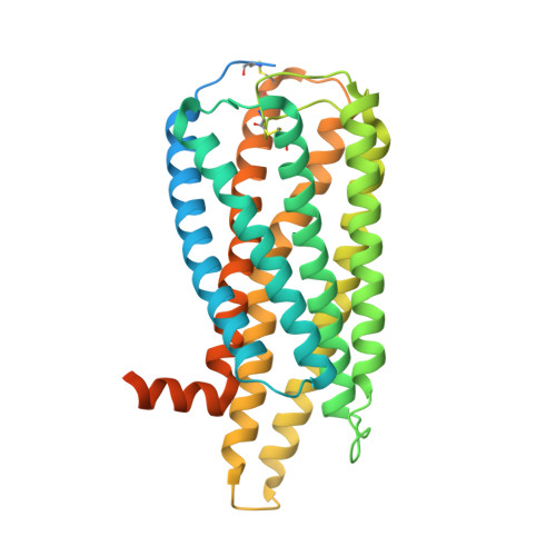
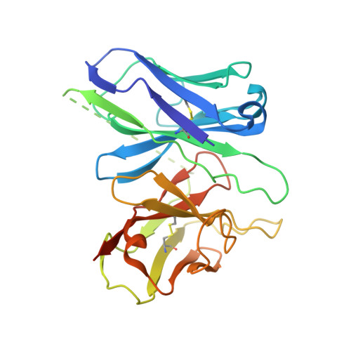
Structural basis for lysophosphatidylserine recognition by GPR34.

Izume, T.Kawahara, R.Uwamizu, A.Chen, L.Yaginuma, S.Omi, J.Kawana, H.Hou, F.Sano, F.K.Tanaka, T.Kobayashi, K.Okamoto, H.H.Kise, Y.Ohwada, T.Aoki, J.Shihoya, W.Nureki, O.

(2024) Nat Commun 15: 902-902

  • DOI: https://doi.org/10.1038/s41467-024-45046-z
  • Primary Citation of Related Structures:  
    8XBE, 8XBG, 8XBH, 8XBI

  • PubMed Abstract: 

    GPR34 is a recently identified G-protein coupled receptor, which has an immunomodulatory role and recognizes lysophosphatidylserine (LysoPS) as a putative ligand. Here, we report cryo-electron microscopy structures of human GPR34-G i complex bound with one of two ligands bound: either the LysoPS analogue S3E-LysoPS, or M1, a derivative of S3E-LysoPS in which oleic acid is substituted with a metabolically stable aromatic fatty acid surrogate. The ligand-binding pocket is laterally open toward the membrane, allowing lateral entry of lipidic agonists into the cavity. The amine and carboxylate groups of the serine moiety are recognized by the charged residue cluster. The acyl chain of S3E-LysoPS is bent and fits into the L-shaped hydrophobic pocket in TM4-5 gap, and the aromatic fatty acid surrogate of M1 fits more appropriately. Molecular dynamics simulations further account for the LysoPS-regioselectivity of GPR34. Thus, using a series of structural and physiological experiments, we provide evidence that chemically unstable 2-acyl LysoPS is the physiological ligand for GPR34. Overall, we anticipate the present structures will pave the way for development of novel anticancer drugs that specifically target GPR34.


  • Organizational Affiliation

    Department of Biological Sciences, Graduate School of Science, The University of Tokyo, Bunkyo-ku, Tokyo, 113-0033, Japan.


Macromolecules
Find similar proteins by:  (by identity cutoff)  |  3D Structure
Entity ID: 1
MoleculeChains Sequence LengthOrganismDetailsImage
Guanine nucleotide-binding protein G(i) subunit alpha-1354Homo sapiensMutation(s): 0 
Gene Names: GNAI1
Membrane Entity: Yes 
UniProt & NIH Common Fund Data Resources
Find proteins for P63096 (Homo sapiens)
Explore P63096 
Go to UniProtKB:  P63096
PHAROS:  P63096
GTEx:  ENSG00000127955 
Entity Groups  
Sequence Clusters30% Identity50% Identity70% Identity90% Identity95% Identity100% Identity
UniProt GroupP63096
Sequence Annotations
Expand
  • Reference Sequence
Find similar proteins by:  (by identity cutoff)  |  3D Structure
Entity ID: 2
MoleculeChains Sequence LengthOrganismDetailsImage
Guanine nucleotide-binding protein G(I)/G(S)/G(T) subunit beta-1344Rattus norvegicusMutation(s): 0 
Gene Names: Gnb1
UniProt
Find proteins for P54311 (Rattus norvegicus)
Explore P54311 
Go to UniProtKB:  P54311
Entity Groups  
Sequence Clusters30% Identity50% Identity70% Identity90% Identity95% Identity100% Identity
UniProt GroupP54311
Sequence Annotations
Expand
  • Reference Sequence
Find similar proteins by:  (by identity cutoff)  |  3D Structure
Entity ID: 3
MoleculeChains Sequence LengthOrganismDetailsImage
Guanine nucleotide-binding protein G(I)/G(S)/G(O) subunit gamma-2C [auth G]68Bos taurusMutation(s): 0 
Gene Names: GNG2
Membrane Entity: Yes 
UniProt
Find proteins for P63212 (Bos taurus)
Explore P63212 
Go to UniProtKB:  P63212
Entity Groups  
Sequence Clusters30% Identity50% Identity70% Identity90% Identity95% Identity100% Identity
UniProt GroupP63212
Sequence Annotations
Expand
  • Reference Sequence
Find similar proteins by:  (by identity cutoff)  |  3D Structure
Entity ID: 4
MoleculeChains Sequence LengthOrganismDetailsImage
Probable G-protein coupled receptor 34D [auth R]396Homo sapiensMutation(s): 0 
Gene Names: GPR34
Membrane Entity: Yes 
UniProt & NIH Common Fund Data Resources
Find proteins for Q9UPC5 (Homo sapiens)
Explore Q9UPC5 
Go to UniProtKB:  Q9UPC5
PHAROS:  Q9UPC5
GTEx:  ENSG00000171659 
Entity Groups  
Sequence Clusters30% Identity50% Identity70% Identity90% Identity95% Identity100% Identity
UniProt GroupQ9UPC5
Sequence Annotations
Expand
  • Reference Sequence
Find similar proteins by:  (by identity cutoff)  |  3D Structure
Entity ID: 5
MoleculeChains Sequence LengthOrganismDetailsImage
scFv16E [auth S]260Mus musculusMutation(s): 0 
Entity Groups  
Sequence Clusters30% Identity50% Identity70% Identity90% Identity95% Identity100% Identity
Sequence Annotations
Expand
  • Reference Sequence
Small Molecules
Ligands 1 Unique
IDChains Name / Formula / InChI Key2D Diagram3D Interactions
KW3 (Subject of Investigation/LOI)
Query on KW3

Download Ideal Coordinates CCD File 
F [auth R](2~{S})-2-azanyl-3-[[(2~{R})-1-ethoxy-3-[3-[2-[(3-phenoxyphenyl)methoxy]phenyl]propanoyloxy]propan-2-yl]oxy-oxidanyl-phosphoryl]oxy-propanoic acid
C30 H36 N O11 P
PWIBLZZVGUIXAU-SXOMAYOGSA-N
Experimental Data & Validation

Experimental Data

  • Method: ELECTRON MICROSCOPY
  • Resolution: 2.83 Å
  • Aggregation State: PARTICLE 
  • Reconstruction Method: SINGLE PARTICLE 

Structure Validation

View Full Validation Report


Entry History & Funding Information

Deposition Data


Funding OrganizationLocationGrant Number
Japan Society for the Promotion of Science (JSPS)Japan21H05037

Revision History  (Full details and data files)

  • Version 1.0: 2023-12-27
    Type: Initial release
  • Version 1.1: 2024-05-15
    Changes: Database references, Source and taxonomy