8X8Q

Structure of enterovirus protease in complex host factor


Experimental Data Snapshot

  • Method: ELECTRON MICROSCOPY
  • Resolution: 3.14 Å
  • Aggregation State: PARTICLE 
  • Reconstruction Method: SINGLE PARTICLE 

wwPDB Validation   3D Report Full Report


This is version 1.0 of the entry. See complete history


Literature

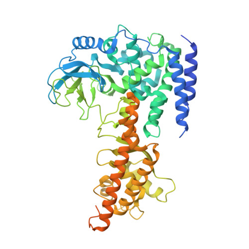
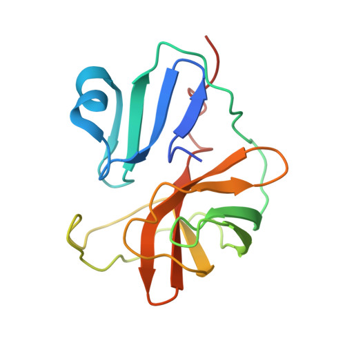
The EV71 2A protease occupies the central cleft of SETD3 and disrupts SETD3-actin interaction.

Gao, X.Wang, B.Zhu, K.Wang, L.Qin, B.Shang, K.Ding, W.Wang, J.Cui, S.

(2024) Nat Commun 15: 4176-4176

  • DOI: https://doi.org/10.1038/s41467-024-48504-w
  • Primary Citation of Related Structures:  
    8X77, 8X8Q

  • PubMed Abstract: 

    SETD3 is an essential host factor for the replication of a variety of enteroviruses that specifically interacts with viral protease 2A. However, the interaction between SETD3 and the 2A protease has not been fully characterized. Here, we use X-ray crystallography and cryo-electron microscopy to determine the structures of SETD3 complexed with the 2A protease of EV71 to 3.5 Å and 3.1 Å resolution, respectively. We find that the 2A protease occupies the V-shaped central cleft of SETD3 through two discrete sites. The relative positions of the two proteins vary in the crystal and cryo-EM structures, showing dynamic binding. A biolayer interferometry assay shows that the EV71 2A protease outcompetes actin for SETD3 binding. We identify key 2A residues involved in SETD3 binding and demonstrate that 2A's ability to bind SETD3 correlates with EV71 production in cells. Coimmunoprecipitation experiments in EV71 infected and 2A expressing cells indicate that 2A interferes with the SETD3-actin complex, and the disruption of this complex reduces enterovirus replication. Together, these results reveal the molecular mechanism underlying the interplay between SETD3, actin, and viral 2A during virus replication.


  • Organizational Affiliation

    NHC Key Laboratory of Systems Biology of Pathogens, National Institute of Pathogen Biology, Chinese Academy of Medical Sciences and Peking Union Medical College, Beijing, 100730, China.


Macromolecules
Find similar proteins by:  (by identity cutoff)  |  3D Structure
Entity ID: 1
MoleculeChains Sequence LengthOrganismDetailsImage
Actin-histidine N-methyltransferaseA [auth B]594Homo sapiensMutation(s): 0 
Gene Names: SETD3
UniProt & NIH Common Fund Data Resources
Find proteins for Q86TU7 (Homo sapiens)
Explore Q86TU7 
Go to UniProtKB:  Q86TU7
PHAROS:  Q86TU7
GTEx:  ENSG00000183576 
Entity Groups  
Sequence Clusters30% Identity50% Identity70% Identity90% Identity95% Identity100% Identity
UniProt GroupQ86TU7
Sequence Annotations
Expand
  • Reference Sequence
Find similar proteins by:  (by identity cutoff)  |  3D Structure
Entity ID: 2
MoleculeChains Sequence LengthOrganismDetailsImage
2A protein (Fragment)B [auth F]150Enterovirus A71Mutation(s): 1 
UniProt
Find proteins for R9YK28 (Human enterovirus 71)
Explore R9YK28 
Go to UniProtKB:  R9YK28
Entity Groups  
Sequence Clusters30% Identity50% Identity70% Identity90% Identity95% Identity100% Identity
UniProt GroupR9YK28
Sequence Annotations
Expand
  • Reference Sequence
Small Molecules
Ligands 1 Unique
IDChains Name / Formula / InChI Key2D Diagram3D Interactions
ZN (Subject of Investigation/LOI)
Query on ZN

Download Ideal Coordinates CCD File 
C [auth F]ZINC ION
Zn
PTFCDOFLOPIGGS-UHFFFAOYSA-N
Experimental Data & Validation

Experimental Data

  • Method: ELECTRON MICROSCOPY
  • Resolution: 3.14 Å
  • Aggregation State: PARTICLE 
  • Reconstruction Method: SINGLE PARTICLE 

Structure Validation

View Full Validation Report


Entry History & Funding Information

Deposition Data


Funding OrganizationLocationGrant Number
Not funded--

Revision History  (Full details and data files)

  • Version 1.0: 2024-05-29
    Type: Initial release