8W3A | pdb_00008w3a

Crystal structure of Shewanella benthica Group 1 truncated hemoglobin C51S C71S variant with trans heme D


Experimental Data Snapshot

  • Method: X-RAY DIFFRACTION
  • Resolution: 1.80 Å
  • R-Value Free: 
    0.236 (Depositor), 0.211 (DCC) 
  • R-Value Work: 
    0.198 (Depositor) 
  • R-Value Observed: 
    0.200 (Depositor) 

Starting Model: experimental
View more details

wwPDB Validation 3D Report Full Report

Validation slider image for 8W3A

Ligand Structure Quality Assessment 


This is version 1.1 of the entry. See complete history

Literature

Heme d formation in a Shewanella benthica hemoglobin.

Martinez Grundman, J.E.Schultz, T.D.Schlessman, J.L.Liu, K.Johnson, E.A.Lecomte, J.T.J.

(2024) J Inorg Biochem 259: 112654-112654

  • DOI: https://doi.org/10.1016/j.jinorgbio.2024.112654
  • Primary Citation Related Structures: 
    8UGZ, 8VSH, 8W3A

  • PubMed Abstract: 

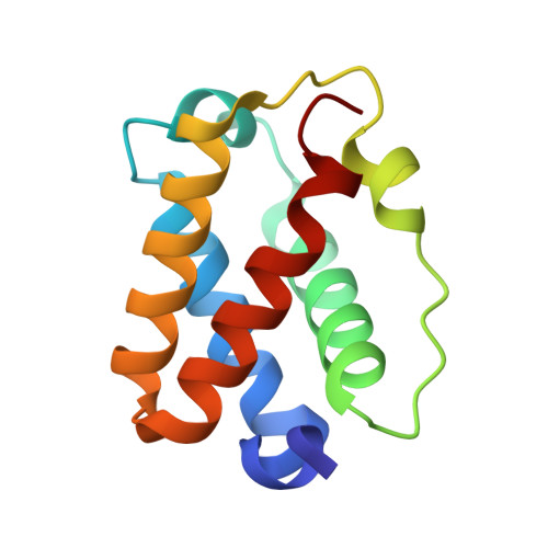
    In our continued investigations of microbial globins, we solved the structure of a truncated hemoglobin from Shewanella benthica, an obligate psychropiezophilic bacterium. The distal side of the heme active site is lined mostly with hydrophobic residues, with the exception of a tyrosine, Tyr34 (CD1) and a histidine, His24 (B13). We found that purified SbHbN, when crystallized in the ferric form with polyethylene glycol as precipitant, turned into a green color over weeks. The electron density obtained from the green crystals accommodated a trans heme d, a chlorin-type derivative featuring a γ-spirolactone and a vicinal hydroxyl group on a pyrroline ring. In solution, exposure of the protein to one equivalent of hydrogen peroxide resulted in a similar green color change, but caused by the formation of multiple products. These were oxidation species released on protein denaturation, likely including heme d, and a species with heme covalently attached to the polypeptide. The Tyr34Phe replacement prevented the formation of both heme d and the covalent linkage. The ready modification of heme b by SbHbN expands the range of chemistries supported by the globin fold and offers a route to a novel heme cofactor.


  • Organizational Affiliation
    • T.C. Jenkins Department of Biophysics, Johns Hopkins University, Baltimore, MD 21218, USA.

Macromolecule Content 

  • Total Structure Weight: 56.35 kDa 
  • Atom Count: 4,261 
  • Modeled Residue Count: 464 
  • Deposited Residue Count: 464 
  • Unique protein chains: 1

Macromolecules

Find similar proteins by:|  3D Structure
Entity ID: 1
MoleculeChains  Sequence LengthOrganismDetailsImage
Group 1 truncated hemoglobin
A, B, C, D
116Shewanella benthica KT99Mutation(s): 2 
Gene Names: KT99_16901
UniProt
Find proteins for A9DF82 (Shewanella benthica KT99)
Explore A9DF82 
Go to UniProtKB:  A9DF82
Entity Groups
Sequence Clusters30% Identity50% Identity70% Identity90% Identity95% Identity100% Identity
UniProt GroupA9DF82
Sequence Annotations
Expand
Reference Sequence

Small Molecules

Ligands 2 Unique
IDChains Name / Formula / InChI Key2D Diagram3D Interactions
A1ADT

Query on A1ADT


Download:Ideal Coordinates CCD File
F [auth A],
G [auth B],
I [auth C],
K [auth D]
{3-[(2R,5'R)-9',14'-diethenyl-5'-hydroxy-5',10',15',19'-tetramethyl-5-oxo-4,5-dihydro-3H-spiro[furan-2,4'-[21,22,23,24]tetraazapentacyclo[16.2.1.13,6.18,11.113,16]tetracosa[1,3(24),6,8,10,12,14,16(22),17,19]decaen]-20'-yl-kappa~4~N~21'~,N~22'~,N~23'~,N~24'~]propanoato}iron
C34 H32 Fe N4 O5
CYBWRCIPTVPWKW-YDXXJHAFSA-M
HEM

Query on HEM


Download:Ideal Coordinates CCD File
E [auth A],
H [auth B],
J [auth C],
L [auth D]
PROTOPORPHYRIN IX CONTAINING FE
C34 H32 Fe N4 O4
KABFMIBPWCXCRK-RGGAHWMASA-L

Experimental Data & Validation

Experimental Data

  • Method: X-RAY DIFFRACTION
  • Resolution: 1.80 Å
  • R-Value Free:  0.236 (Depositor), 0.211 (DCC) 
  • R-Value Work:  0.198 (Depositor) 
  • R-Value Observed: 0.200 (Depositor) 
Space Group: P 21 21 21
Unit Cell:
Length ( Å )Angle ( ˚ )
a = 27.357α = 90
b = 104.809β = 90
c = 150.551γ = 90
Software Package:
Software NamePurpose
PHENIXrefinement
CrysalisProdata scaling
CrysalisProdata reduction
PHASERphasing

Structure Validation

View Full Validation Report


Ligand Structure Quality Assessment 


Entry History 

& Funding Information

Deposition Data


Funding OrganizationLocationGrant Number
National Science Foundation (NSF, United States)United StatesCHE-2003950

Revision History  (Full details and data files)

  • Version 1.0: 2024-04-03
    Type: Initial release
  • Version 1.1: 2024-07-17
    Changes: Database references