8VOL

Apex domain deletion mutant of bacteriophage P2 central spike protein, membrane-piercing module


Experimental Data Snapshot

  • Method: X-RAY DIFFRACTION
  • Resolution: 0.90 Å
  • R-Value Free: 0.127 
  • R-Value Work: 0.116 
  • R-Value Observed: 0.116 

Starting Model: experimental
View more details

wwPDB Validation   3D Report Full Report


Ligand Structure Quality Assessment 


This is version 1.0 of the entry. See complete history


Literature

Function of the bacteriophage P2 baseplate central spike Apex domain in the infection process.

Miller, J.M.Knyazhanskaya, E.S.Buth, S.A.Prokhorov, N.S.Leiman, P.G.

(2023) bioRxiv 

  • DOI: https://doi.org/10.1101/2023.02.25.529910
  • Primary Citation of Related Structures:  
    8VOL, 8VOM, 8VON

  • PubMed Abstract: 

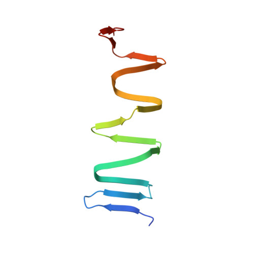
    The contractile tail of bacteriophage P2 functions to drive the tail tube across the outer membrane of its host bacterium, a prerequisite event for subsequent translocation of phage genomic DNA into the host cell. The tube is equipped with a spike-shaped protein (product of P2 gene V , gpV or Spike) that contains a membrane-attacking Apex domain carrying a centrally positioned Fe ion. The ion is enclosed in a histidine cage that is formed by three symmetry-related copies of a conserved HxH (histidine, any residue, histidine) sequence motif. Here, we used solution biophysics and X-ray crystallography to characterize the structure and properties of Spike mutants in which the Apex domain was either deleted or its histidine cage was either destroyed or replaced with a hydrophobic core. We found that the Apex domain is not required for the folding of full-length gpV or its middle intertwined β-helical domain. Furthermore, despite its high conservation, the Apex domain is dispensable for infection in laboratory conditions. Collectively, our results show that the diameter of the Spike but not the nature of its Apex domain determines the efficiency of infection, which further strengthens the earlier hypothesis of a drill bit-like function of the Spike in host envelope disruption.


Macromolecules
Find similar proteins by:  (by identity cutoff)  |  3D Structure
Entity ID: 1
MoleculeChains Sequence LengthOrganismDetailsImage
Spike protein
A, B, C, D, E
A, B, C, D, E, F
104Bacteriophage P2Mutation(s): 1 
UniProt
Find proteins for P31340 (Escherichia phage P2)
Explore P31340 
Go to UniProtKB:  P31340
Entity Groups  
Sequence Clusters30% Identity50% Identity70% Identity90% Identity95% Identity100% Identity
UniProt GroupP31340
Sequence Annotations
Expand
  • Reference Sequence
Small Molecules
Ligands 1 Unique
IDChains Name / Formula / InChI Key2D Diagram3D Interactions
PTY
Query on PTY

Download Ideal Coordinates CCD File 
G [auth D]PHOSPHATIDYLETHANOLAMINE
C40 H80 N O8 P
NJGIRBISCGPRPF-KXQOOQHDSA-N
Experimental Data & Validation

Experimental Data

  • Method: X-RAY DIFFRACTION
  • Resolution: 0.90 Å
  • R-Value Free: 0.127 
  • R-Value Work: 0.116 
  • R-Value Observed: 0.116 
  • Space Group: P 1
Unit Cell:
Length ( Å )Angle ( ˚ )
a = 45.52α = 91.61
b = 49.91β = 88.25
c = 72.93γ = 111.65
Software Package:
Software NamePurpose
XDSdata reduction
XSCALEdata scaling
PHASERphasing
Cootmodel building
PHENIXrefinement

Structure Validation

View Full Validation Report


Ligand Structure Quality Assessment 


Entry History & Funding Information

Deposition Data


Funding OrganizationLocationGrant Number
National Institutes of Health/National Institute of General Medical Sciences (NIH/NIGMS)United StatesGM139034

Revision History  (Full details and data files)

  • Version 1.0: 2024-08-07
    Type: Initial release