8VHF | pdb_00008vhf

Cryo-EM structure of GPR119-Gs-Nb35 complex with small molecule agonist MBX-2982


Experimental Data Snapshot

  • Method: ELECTRON MICROSCOPY
  • Resolution: 3.51 Å
  • Aggregation State: PARTICLE 
  • Reconstruction Method: SINGLE PARTICLE 

Starting Model: experimental
View more details

wwPDB Validation 3D Report Full Report

Validation slider image for 8VHF

This is version 1.2 of the entry. See complete history

Literature

Native mass spectrometry prescreening of G protein-coupled receptor complexes for cryo-EM structure determination.

Kim, D.Liu, W.Viner, R.Cherezov, V.

(2024) Structure 32: 2206-2219.e4

  • DOI: https://doi.org/10.1016/j.str.2024.10.004
  • Primary Citation Related Structures: 
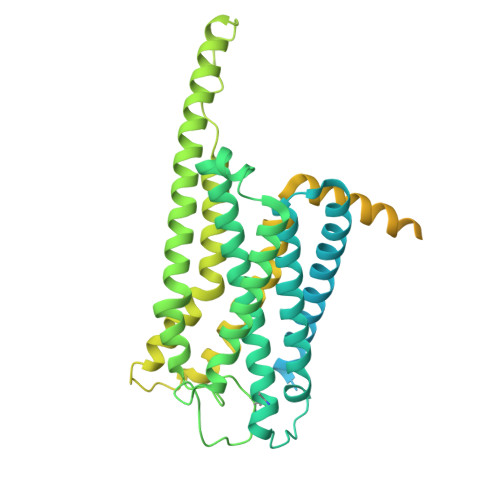
    8VHF

  • PubMed Abstract: 

    G protein-coupled receptors (GPCRs) are essential transmembrane proteins playing key roles in human health and disease. Understanding their atomic-level molecular structure and conformational states is imperative for advancing drug development. Recent breakthroughs in single-particle cryogenic electron microscopy (cryo-EM) have propelled the structural biology of GPCRs into a new era. Nevertheless, the preparation of suitable GPCR samples and their complexes for cryo-EM analysis remains challenging due to their poor stability and highly dynamic nature. Here, we present our online buffer exchange-native MS method combined with Direct Mass Technology (OBE-nMS+DMT) which facilitates high-throughput analysis and guides sample preparation. We applied this method to optimize the GPR119-G s complex sample prior to cryo-EM analysis, leading to a 3.51 Å resolution structure from only 396 movies collected on a 200 kV Glacios. This study suggests that the OBE-nMS+DMT method emerges as a powerful tool for prescreening sample conditions in cryo-EM studies of GPCRs and other membrane protein complexes.


  • Organizational Affiliation
    • Bridge Institute, Michelson Center for Convergent Bioscience, University of Southern California, Los Angeles, CA 90089, USA; Department of Chemistry, University of Southern California, Los Angeles, CA 90089, USA.

Macromolecule Content 

  • Total Structure Weight: 176.95 kDa 
  • Atom Count: 8,299 
  • Modeled Residue Count: 1,057 
  • Deposited Residue Count: 1,589 
  • Unique protein chains: 5

Macromolecules

Find similar proteins by:|  3D Structure
Entity ID: 1
MoleculeChains  Sequence LengthOrganismDetailsImage
Guanine nucleotide-binding protein G(I)/G(S)/G(T) subunit beta-1, HiBiTA [auth B]371Homo sapiensMutation(s): 0 
Gene Names: GNB1
UniProt & NIH Common Fund Data Resources
Find proteins for P62873 (Homo sapiens)
Explore P62873 
Go to UniProtKB:  P62873
PHAROS:  P62873
GTEx:  ENSG00000078369 
Entity Groups
Sequence Clusters30% Identity50% Identity70% Identity90% Identity95% Identity100% Identity
UniProt GroupP62873
Sequence Annotations
Expand
Reference Sequence
Find similar proteins by:|  3D Structure
Entity ID: 2
MoleculeChains  Sequence LengthOrganismDetailsImage
Guanine nucleotide-binding protein G(I)/G(S)/G(O) subunit gamma-2B [auth C]71Homo sapiensMutation(s): 0 
Gene Names: GNG2
Membrane Entity: Yes 
UniProt & NIH Common Fund Data Resources
Find proteins for P59768 (Homo sapiens)
Explore P59768 
Go to UniProtKB:  P59768
PHAROS:  P59768
GTEx:  ENSG00000186469 
Entity Groups
Sequence Clusters30% Identity50% Identity70% Identity90% Identity95% Identity100% Identity
UniProt GroupP59768
Sequence Annotations
Expand
Reference Sequence
Find similar proteins by:|  3D Structure
Entity ID: 3
MoleculeChains  Sequence LengthOrganismDetailsImage
Nanobody35C [auth N]138Lama glamaMutation(s): 0 
Find similar proteins by:|  3D Structure
Entity ID: 4
MoleculeChains  Sequence LengthOrganismDetailsImage
Guanine nucleotide-binding protein G(s) subunit alpha isoforms shortD [auth A]394Homo sapiensMutation(s): 8 
Gene Names: GNASGNAS1GSP
EC: 3.6.5
Membrane Entity: Yes 
UniProt & NIH Common Fund Data Resources
Find proteins for P63092 (Homo sapiens)
Explore P63092 
Go to UniProtKB:  P63092
PHAROS:  P63092
GTEx:  ENSG00000087460 
Entity Groups
Sequence Clusters30% Identity50% Identity70% Identity90% Identity95% Identity100% Identity
UniProt GroupP63092
Sequence Annotations
Expand
Reference Sequence
Find similar proteins by:|  3D Structure
Entity ID: 5
MoleculeChains  Sequence LengthOrganismDetailsImage
Soluble cytochrome b562,Glucose-dependent insulinotropic receptor,LgBiTE [auth R]615Escherichia coliHomo sapiensOplophorus gracilirostris
This entity is chimeric
Mutation(s): 3 
Gene Names: cybCGPR119
Membrane Entity: Yes 
UniProt & NIH Common Fund Data Resources
Find proteins for P0ABE7 (Escherichia coli)
Explore P0ABE7 
Go to UniProtKB:  P0ABE7
Find proteins for Q8TDV5 (Homo sapiens)
Explore Q8TDV5 
Go to UniProtKB:  Q8TDV5
PHAROS:  Q8TDV5
GTEx:  ENSG00000147262 
Entity Groups
Sequence Clusters30% Identity50% Identity70% Identity90% Identity95% Identity100% Identity
UniProt GroupsQ8TDV5P0ABE7
Sequence Annotations
Expand
Reference Sequence

Small Molecules

Ligands 1 Unique
IDChains Name / Formula / InChI Key2D Diagram3D Interactions
8VP
(Subject of Investigation/LOI)

Query on 8VP


Download:Ideal Coordinates CCD File
F [auth R]2-[1-(5-ethylpyrimidin-2-yl)piperidin-4-yl]-4-[[4-(1,2,3,4-tetrazol-1-yl)phenoxy]methyl]-1,3-thiazole
C22 H24 N8 O S
NFTMKHWBOINJGM-UHFFFAOYSA-N

Experimental Data & Validation

Experimental Data

  • Method: ELECTRON MICROSCOPY
  • Resolution: 3.51 Å
  • Aggregation State: PARTICLE 
  • Reconstruction Method: SINGLE PARTICLE 
EM Software:
TaskSoftware PackageVersion
RECONSTRUCTIONcryoSPARC4.3.1
MODEL REFINEMENTPHENIX1.21rc1_5156:

Structure Validation

View Full Validation Report


Entry History 

& Funding Information

Deposition Data


Funding OrganizationLocationGrant Number
National Institutes of Health/National Institute of General Medical Sciences (NIH/NIGMS)United StatesGM127086

Revision History  (Full details and data files)

  • Version 1.0: 2024-10-30
    Type: Initial release
  • Version 1.1: 2024-11-13
    Changes: Data collection
  • Version 1.2: 2025-03-05
    Changes: Data collection, Database references