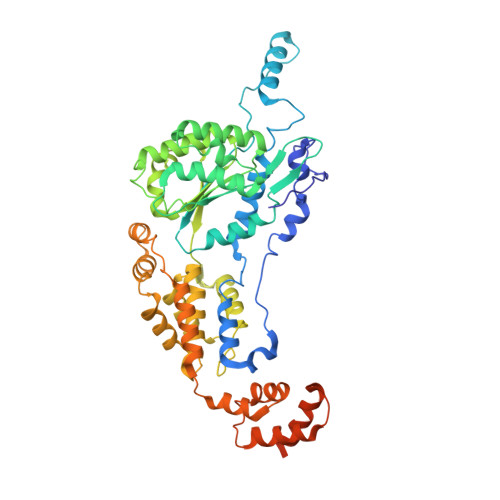
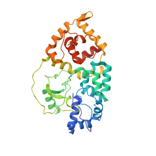
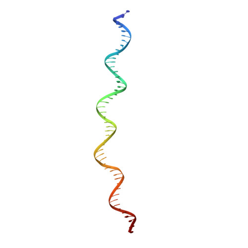
8VCJ

CryoEM structure of the TnsC(1-503)-TnsD(1-318)-DNA complex in a 7:2:1 stoichiometry from E. coli Tn7 bound to ATPgS and ADP


Experimental Data Snapshot

  • Method: ELECTRON MICROSCOPY
  • Resolution: 3.32 Å
  • Aggregation State: PARTICLE 
  • Reconstruction Method: SINGLE PARTICLE 

Starting Model: experimental
View more details

wwPDB Validation   3D Report Full Report


This is version 1.0 of the entry. See complete history


Literature

Assembly of the Tn7 targeting complex by a regulated stepwise process.

Shen, Y.Krishnan, S.S.Petassi, M.T.Hancock, M.A.Peters, J.E.Guarne, A.

(2024) Mol Cell 84: 2368-2381.e6

  • DOI: https://doi.org/10.1016/j.molcel.2024.05.012

  • PubMed Abstract: 

    The Tn7 family of transposons is notable for its highly regulated integration mechanisms, including programmable RNA-guided transposition. The targeting pathways rely on dedicated target selection proteins from the TniQ family and the AAA+ adaptor TnsC to recruit and activate the transposase at specific target sites. Here, we report the cryoelectron microscopy (cryo-EM) structures of TnsC bound to the TniQ domain of TnsD from prototypical Tn7 and unveil key regulatory steps stemming from unique behaviors of ATP- versus ADP-bound TnsC. We show that TnsD recruits ADP-bound dimers of TnsC and acts as an exchange factor to release one protomer with exchange to ATP. This loading process explains how TnsC assembles a heptameric ring unidirectionally from the target site. This unique loading process results in functionally distinct TnsC protomers within the ring, providing a checkpoint for target immunity and explaining how insertions at programmed sites precisely occur in a specific orientation across Tn7 elements.


  • Organizational Affiliation

    Department of Biochemistry, McGill University, Montreal, QC H3G 0B1, Canada; Centre de recherche en biologie structurale (CRBS), McGill University, Montreal, QC H3G 0B1, Canada.


Macromolecules

Find similar proteins by:  (by identity cutoff)  |  3D Structure
Entity ID: 1
MoleculeChains Sequence LengthOrganismDetailsImage
Transposon Tn7 transposition protein TnsC523Escherichia coliMutation(s): 1 
Gene Names: tnsC
UniProt
Find proteins for P05846 (Escherichia coli)
Explore P05846 
Go to UniProtKB:  P05846
Entity Groups  
Sequence Clusters30% Identity50% Identity70% Identity90% Identity95% Identity100% Identity
UniProt GroupP05846
Sequence Annotations
Expand
  • Reference Sequence
Find similar proteins by:  (by identity cutoff)  |  3D Structure
Entity ID: 2
MoleculeChains Sequence LengthOrganismDetailsImage
Transposon Tn7 transposition protein TnsDD [auth Y],
I [auth X]
318Escherichia coliMutation(s): 0 
Gene Names: tnsD
UniProt
Find proteins for P13991 (Escherichia coli)
Explore P13991 
Go to UniProtKB:  P13991
Entity Groups  
Sequence Clusters30% Identity50% Identity70% Identity90% Identity95% Identity100% Identity
UniProt GroupP13991
Sequence Annotations
Expand
  • Reference Sequence
Find similar nucleic acids by:  (by identity cutoff)  |  3D Structure
Entity ID: 3
MoleculeChains LengthOrganismImage
DNAJ [auth H]50Escherichia coli
Sequence Annotations
Expand
  • Reference Sequence
Find similar nucleic acids by:  (by identity cutoff)  |  3D Structure
Entity ID: 4
MoleculeChains LengthOrganismImage
DNAK [auth I]50Escherichia coli
Sequence Annotations
Expand
  • Reference Sequence
Small Molecules
Ligands 4 Unique
IDChains Name / Formula / InChI Key2D Diagram3D Interactions
AGS (Subject of Investigation/LOI)
Query on AGS

Download Ideal Coordinates CCD File 
P [auth C],
Y [auth G]
PHOSPHOTHIOPHOSPHORIC ACID-ADENYLATE ESTER
C10 H16 N5 O12 P3 S
NLTUCYMLOPLUHL-KQYNXXCUSA-N
ADP (Subject of Investigation/LOI)
Query on ADP

Download Ideal Coordinates CCD File 
L [auth A],
N [auth B],
S [auth D],
U [auth E],
X [auth F]
ADENOSINE-5'-DIPHOSPHATE
C10 H15 N5 O10 P2
XTWYTFMLZFPYCI-KQYNXXCUSA-N
ZN (Subject of Investigation/LOI)
Query on ZN

Download Ideal Coordinates CCD File 
AA [auth X],
R [auth Y]
ZINC ION
Zn
PTFCDOFLOPIGGS-UHFFFAOYSA-N
MG (Subject of Investigation/LOI)
Query on MG

Download Ideal Coordinates CCD File 
M [auth A]
O [auth B]
Q [auth C]
T [auth D]
V [auth E]
M [auth A],
O [auth B],
Q [auth C],
T [auth D],
V [auth E],
W [auth E],
Z [auth G]
MAGNESIUM ION
Mg
JLVVSXFLKOJNIY-UHFFFAOYSA-N
Experimental Data & Validation

Experimental Data

  • Method: ELECTRON MICROSCOPY
  • Resolution: 3.32 Å
  • Aggregation State: PARTICLE 
  • Reconstruction Method: SINGLE PARTICLE 
EM Software:
TaskSoftware PackageVersion
MODEL REFINEMENTPHENIX1.19.2_4158:

Structure Validation

View Full Validation Report


Entry History & Funding Information

Deposition Data


Funding OrganizationLocationGrant Number
Canadian Institutes of Health Research (CIHR)CanadaPJT-155941

Revision History  (Full details and data files)

  • Version 1.0: 2024-07-31
    Type: Initial release