8GLU | pdb_00008glu

CryoEM structure of TnsC(1-503) bound to TnsD(1-318) from E.coli Tn7


Experimental Data Snapshot

  • Method: ELECTRON MICROSCOPY
  • Resolution: 3.57 Å
  • Aggregation State: PARTICLE 
  • Reconstruction Method: SINGLE PARTICLE 

Starting Models: experimental, other
View more details

wwPDB Validation 3D Report Full Report

Validation slider image for 8GLU

This is version 1.0 of the entry. See complete history

Literature

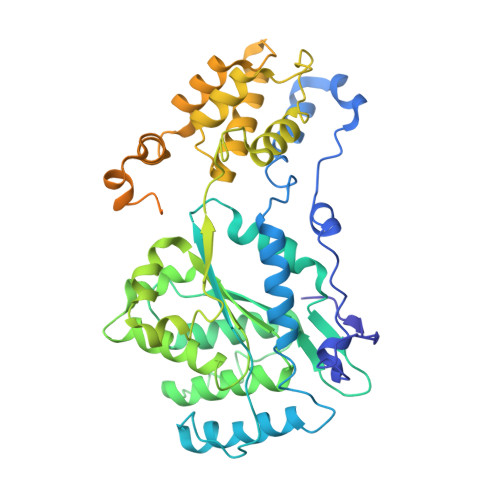
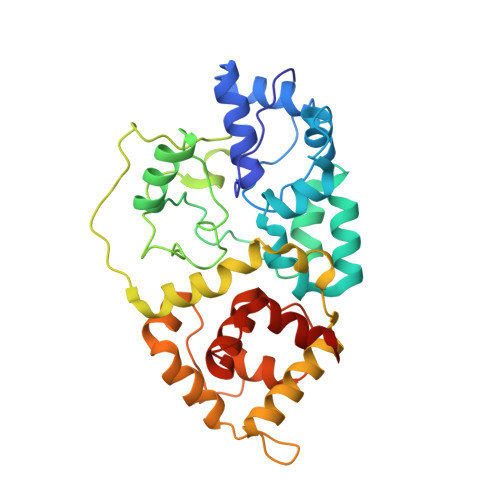
Assembly of the Tn7 targeting complex by a regulated stepwise process.

Shen, Y.Krishnan, S.S.Petassi, M.T.Hancock, M.A.Peters, J.E.Guarne, A.

(2024) Mol Cell 84: 2368-2381.e6

  • DOI: https://doi.org/10.1016/j.molcel.2024.05.012
  • Primary Citation Related Structures: 
    8GLU, 8GLW, 8GLX, 8VCJ, 8VCT

  • PubMed Abstract: 

    The Tn7 family of transposons is notable for its highly regulated integration mechanisms, including programmable RNA-guided transposition. The targeting pathways rely on dedicated target selection proteins from the TniQ family and the AAA+ adaptor TnsC to recruit and activate the transposase at specific target sites. Here, we report the cryoelectron microscopy (cryo-EM) structures of TnsC bound to the TniQ domain of TnsD from prototypical Tn7 and unveil key regulatory steps stemming from unique behaviors of ATP- versus ADP-bound TnsC. We show that TnsD recruits ADP-bound dimers of TnsC and acts as an exchange factor to release one protomer with exchange to ATP. This loading process explains how TnsC assembles a heptameric ring unidirectionally from the target site. This unique loading process results in functionally distinct TnsC protomers within the ring, providing a checkpoint for target immunity and explaining how insertions at programmed sites precisely occur in a specific orientation across Tn7 elements.


  • Organizational Affiliation
    • Department of Biochemistry, McGill University, Montreal, QC H3G 0B1, Canada; Centre de recherche en biologie structurale (CRBS), McGill University, Montreal, QC H3G 0B1, Canada.

Macromolecule Content 

  • Total Structure Weight: 216.17 kDa 
  • Atom Count: 12,338 
  • Modeled Residue Count: 1,533 
  • Deposited Residue Count: 1,887 
  • Unique protein chains: 2

Macromolecules

Find similar proteins by:|  3D Structure
Entity ID: 1
MoleculeChains  Sequence LengthOrganismDetailsImage
Transposon Tn7 transposition protein TnsCA [auth G],
C [auth F],
D [auth E]
523Escherichia coliMutation(s): 1 
Gene Names: tnsC
UniProt
Find proteins for P05846 (Escherichia coli)
Explore P05846 
Go to UniProtKB:  P05846
Entity Groups
Sequence Clusters30% Identity50% Identity70% Identity90% Identity95% Identity100% Identity
UniProt GroupP05846
Sequence Annotations
Expand
Reference Sequence
Find similar proteins by:|  3D Structure
Entity ID: 2
MoleculeChains  Sequence LengthOrganismDetailsImage
Transposon Tn7 transposition protein TnsDB [auth X]318Escherichia coliMutation(s): 0 
Gene Names: tnsD
UniProt
Find proteins for P13991 (Escherichia coli)
Explore P13991 
Go to UniProtKB:  P13991
Entity Groups
Sequence Clusters30% Identity50% Identity70% Identity90% Identity95% Identity100% Identity
UniProt GroupP13991
Sequence Annotations
Expand
Reference Sequence

Small Molecules

Ligands 4 Unique
IDChains Name / Formula / InChI Key2D Diagram3D Interactions
ATP
(Subject of Investigation/LOI)

Query on ATP


Download:Ideal Coordinates CCD File
E [auth G],
H [auth F]
ADENOSINE-5'-TRIPHOSPHATE
C10 H16 N5 O13 P3
ZKHQWZAMYRWXGA-KQYNXXCUSA-N
ADP
(Subject of Investigation/LOI)

Query on ADP


Download:Ideal Coordinates CCD File
J [auth E]ADENOSINE-5'-DIPHOSPHATE
C10 H15 N5 O10 P2
XTWYTFMLZFPYCI-KQYNXXCUSA-N
ZN
(Subject of Investigation/LOI)

Query on ZN


Download:Ideal Coordinates CCD File
G [auth X]ZINC ION
Zn
PTFCDOFLOPIGGS-UHFFFAOYSA-N
MG
(Subject of Investigation/LOI)

Query on MG


Download:Ideal Coordinates CCD File
F [auth G],
I [auth F],
K [auth E]
MAGNESIUM ION
Mg
JLVVSXFLKOJNIY-UHFFFAOYSA-N

Experimental Data & Validation

Experimental Data

  • Method: ELECTRON MICROSCOPY
  • Resolution: 3.57 Å
  • Aggregation State: PARTICLE 
  • Reconstruction Method: SINGLE PARTICLE 
EM Software:
TaskSoftware PackageVersion
MODEL REFINEMENTPHENIX1.19.2_4158:

Structure Validation

View Full Validation Report


Entry History 

& Funding Information

Deposition Data


Funding OrganizationLocationGrant Number
Canadian Institutes of Health Research (CIHR)CanadaPJT-155941

Revision History  (Full details and data files)

  • Version 1.0: 2024-07-31
    Type: Initial release