8UXI

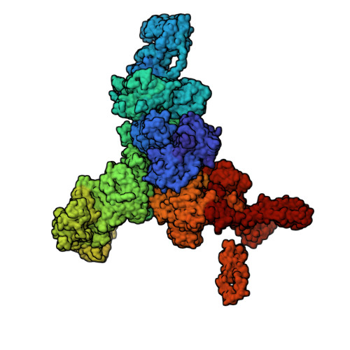
Structure of PKA phosphorylated human RyR2-R420W in the open state in the presence of calcium


Experimental Data Snapshot

  • Method: ELECTRON MICROSCOPY
  • Resolution: 3.29 Å
  • Aggregation State: PARTICLE 
  • Reconstruction Method: SINGLE PARTICLE 

Starting Model: experimental
View more details

wwPDB Validation   3D Report Full Report


This is version 1.2 of the entry. See complete history


Literature

Structural basis for ryanodine receptor type 2 leak in heart failure and arrhythmogenic disorders.

Miotto, M.C.Reiken, S.Wronska, A.Yuan, Q.Dridi, H.Liu, Y.Weninger, G.Tchagou, C.Marks, A.R.

(2024) Nat Commun 15: 8080-8080

  • DOI: https://doi.org/10.1038/s41467-024-51791-y
  • Primary Citation of Related Structures:  
    8UQ2, 8UQ3, 8UQ4, 8UQ5, 8UXC, 8UXE, 8UXF, 8UXG, 8UXH, 8UXI, 8UXL, 8UXM

  • PubMed Abstract: 

    Heart failure, the leading cause of mortality and morbidity in the developed world, is characterized by cardiac ryanodine receptor 2 channels that are hyperphosphorylated, oxidized, and depleted of the stabilizing subunit calstabin-2. This results in a diastolic sarcoplasmic reticulum Ca 2+ leak that impairs cardiac contractility and triggers arrhythmias. Genetic mutations in ryanodine receptor 2 can also cause Ca 2+ leak, leading to arrhythmias and sudden cardiac death. Here, we solved the cryogenic electron microscopy structures of ryanodine receptor 2 variants linked either to heart failure or inherited sudden cardiac death. All are in the primed state, part way between closed and open. Binding of Rycal drugs to ryanodine receptor 2 channels reverts the primed state back towards the closed state, decreasing Ca 2+ leak, improving cardiac function, and preventing arrhythmias. We propose a structural-physiological mechanism whereby the ryanodine receptor 2 channel primed state underlies the arrhythmias in heart failure and arrhythmogenic disorders.


  • Organizational Affiliation

    Department of Physiology and Cellular Biophysics, Columbia University Vagelos College of Physicians and Surgeons, New York, NY, USA. mm5642@cumc.columbia.edu.


Macromolecules
Find similar proteins by:  (by identity cutoff)  |  3D Structure
Entity ID: 1
MoleculeChains Sequence LengthOrganismDetailsImage
Ryanodine receptor 2A,
F [auth B],
G [auth C],
H [auth D]
4,967Homo sapiensMutation(s): 1 
Gene Names: RYR2
UniProt & NIH Common Fund Data Resources
Find proteins for Q92736 (Homo sapiens)
Explore Q92736 
Go to UniProtKB:  Q92736
PHAROS:  Q92736
GTEx:  ENSG00000198626 
Entity Groups  
Sequence Clusters30% Identity50% Identity70% Identity90% Identity95% Identity100% Identity
UniProt GroupQ92736
Sequence Annotations
Expand
  • Reference Sequence
Find similar proteins by:  (by identity cutoff)  |  3D Structure
Entity ID: 2
MoleculeChains Sequence LengthOrganismDetailsImage
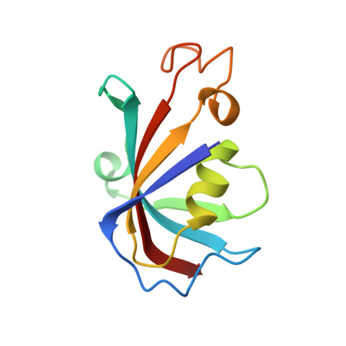
Peptidyl-prolyl cis-trans isomerase FKBP1BB [auth E],
C [auth F],
D [auth G],
E [auth H]
108Homo sapiensMutation(s): 0 
Gene Names: FKBP1BFKBP12.6FKBP1LFKBP9OTK4
EC: 5.2.1.8
UniProt & NIH Common Fund Data Resources
Find proteins for P68106 (Homo sapiens)
Explore P68106 
Go to UniProtKB:  P68106
PHAROS:  P68106
GTEx:  ENSG00000119782 
Entity Groups  
Sequence Clusters30% Identity50% Identity70% Identity90% Identity95% Identity100% Identity
UniProt GroupP68106
Sequence Annotations
Expand
  • Reference Sequence
Small Molecules
Ligands 3 Unique
IDChains Name / Formula / InChI Key2D Diagram3D Interactions
ATP (Subject of Investigation/LOI)
Query on ATP

Download Ideal Coordinates CCD File 
J [auth A]
L [auth A]
N [auth B]
P [auth B]
R [auth C]
J [auth A],
L [auth A],
N [auth B],
P [auth B],
R [auth C],
T [auth C],
V [auth D],
X [auth D]
ADENOSINE-5'-TRIPHOSPHATE
C10 H16 N5 O13 P3
ZKHQWZAMYRWXGA-KQYNXXCUSA-N
ZN (Subject of Investigation/LOI)
Query on ZN

Download Ideal Coordinates CCD File 
I [auth A],
M [auth B],
Q [auth C],
U [auth D]
ZINC ION
Zn
PTFCDOFLOPIGGS-UHFFFAOYSA-N
CA (Subject of Investigation/LOI)
Query on CA

Download Ideal Coordinates CCD File 
K [auth A],
O [auth B],
S [auth C],
W [auth D]
CALCIUM ION
Ca
BHPQYMZQTOCNFJ-UHFFFAOYSA-N
Experimental Data & Validation

Experimental Data

  • Method: ELECTRON MICROSCOPY
  • Resolution: 3.29 Å
  • Aggregation State: PARTICLE 
  • Reconstruction Method: SINGLE PARTICLE 
EM Software:
TaskSoftware PackageVersion
MODEL REFINEMENTPHENIX
RECONSTRUCTIONcryoSPARC

Structure Validation

View Full Validation Report


Entry History & Funding Information

Deposition Data


Funding OrganizationLocationGrant Number
National Institutes of Health/National Heart, Lung, and Blood Institute (NIH/NHLBI)United StatesR01HL145473

Revision History  (Full details and data files)

  • Version 1.0: 2023-11-22
    Type: Initial release
  • Version 1.1: 2024-10-02
    Changes: Data collection, Database references
  • Version 1.2: 2024-11-13
    Changes: Data collection, Structure summary