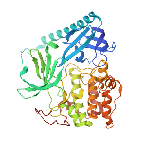
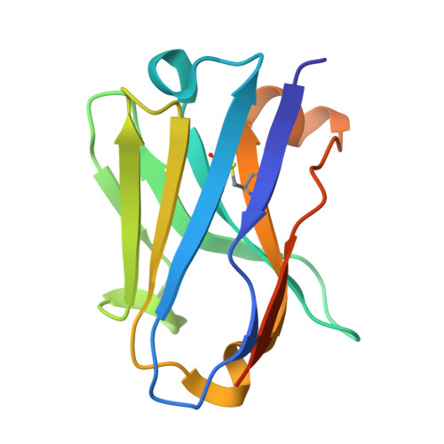
8UW2

Structure of AKT1(E17K) with compound 3 (zinc-free)


Experimental Data Snapshot

  • Method: X-RAY DIFFRACTION
  • Resolution: 2.20 Å
  • R-Value Free: 0.279 
  • R-Value Work: 0.220 
  • R-Value Observed: 0.223 

Starting Model: experimental
View more details

wwPDB Validation   3D Report Full Report


Ligand Structure Quality Assessment 


This is version 1.2 of the entry. See complete history


Literature

Mutant-selective AKT inhibition through lysine targeting and neo-zinc chelation.

Craven, G.B.Chu, H.Sun, J.D.Carelli, J.D.Coyne, B.Chen, H.Chen, Y.Ma, X.Das, S.Kong, W.Zajdlik, A.D.Yang, K.S.Reisberg, S.H.Thompson, P.A.Lipford, J.R.Taunton, J.

(2024) Nature 

  • DOI: https://doi.org/10.1038/s41586-024-08176-4
  • Primary Citation of Related Structures:  
    8UVY, 8UW2, 8UW7, 8UW9, 9C1W

  • PubMed Abstract: 

    Somatic alterations in the oncogenic kinase AKT1 have been identified in a broad spectrum of solid tumours. The most common AKT1 alteration replaces Glu17 with Lys (E17K) in the regulatory pleckstrin homology domain 1 , resulting in constitutive membrane localization and activation of oncogenic signalling. In clinical studies, pan-AKT inhibitors have been found to cause dose-limiting hyperglycaemia 2-6 , which has motivated the search for mutant-selective inhibitors. We exploited the E17K mutation to design allosteric, lysine-targeted salicylaldehyde inhibitors with selectivity for AKT1 (E17K) over wild-type AKT paralogues, a major challenge given the presence of three conserved lysines near the allosteric site. Crystallographic analysis of the covalent inhibitor complex unexpectedly revealed an adventitious tetrahedral zinc ion that coordinates two proximal cysteines in the kinase activation loop while simultaneously engaging the E17K-imine conjugate. The salicylaldimine complex with AKT1 (E17K), but not that with wild-type AKT1, recruits endogenous Zn 2+ in cells, resulting in sustained inhibition. A salicylaldehyde-based inhibitor was efficacious in AKT1 (E17K) tumour xenograft models at doses that did not induce hyperglycaemia. Our study demonstrates the potential to achieve exquisite residence-time-based selectivity for AKT1 (E17K) by targeting the mutant lysine together with Zn 2+ chelation by the resulting salicylaldimine adduct.


  • Organizational Affiliation

    Department of Cellular and Molecular Pharmacology, University of California, San Francisco, San Francisco, CA, USA.


Macromolecules
Find similar proteins by:  (by identity cutoff)  |  3D Structure
Entity ID: 1
MoleculeChains Sequence LengthOrganismDetailsImage
RAC-alpha serine/threonine-protein kinase438Homo sapiensMutation(s): 0 
Gene Names: AKT1
EC: 2.7.11.1
UniProt & NIH Common Fund Data Resources
Find proteins for P31749 (Homo sapiens)
Explore P31749 
Go to UniProtKB:  P31749
PHAROS:  P31749
GTEx:  ENSG00000142208 
Entity Groups  
Sequence Clusters30% Identity50% Identity70% Identity90% Identity95% Identity100% Identity
UniProt GroupP31749
Sequence Annotations
Expand
  • Reference Sequence
Find similar proteins by:  (by identity cutoff)  |  3D Structure
Entity ID: 2
MoleculeChains Sequence LengthOrganismDetailsImage
NB41126Lama glamaMutation(s): 0 
Entity Groups  
Sequence Clusters30% Identity50% Identity70% Identity90% Identity95% Identity100% Identity
Sequence Annotations
Expand
  • Reference Sequence
Small Molecules
Ligands 3 Unique
IDChains Name / Formula / InChI Key2D Diagram3D Interactions
XOO (Subject of Investigation/LOI)
Query on XOO

Download Ideal Coordinates CCD File 
D [auth A]4-{2-[({4-[(2P)-2-(2-aminopyridin-3-yl)-5-phenyl-3H-imidazo[4,5-b]pyridin-3-yl]phenyl}methyl)amino]ethyl}-2-hydroxybenzaldehyde
C33 H28 N6 O2
OMBAVJOHMDVMKX-UHFFFAOYSA-N
SO4
Query on SO4

Download Ideal Coordinates CCD File 
F [auth A]
G [auth A]
H [auth A]
I [auth A]
J [auth A]
F [auth A],
G [auth A],
H [auth A],
I [auth A],
J [auth A],
K [auth B]
SULFATE ION
O4 S
QAOWNCQODCNURD-UHFFFAOYSA-L
EDO
Query on EDO

Download Ideal Coordinates CCD File 
C [auth A],
E [auth A]
1,2-ETHANEDIOL
C2 H6 O2
LYCAIKOWRPUZTN-UHFFFAOYSA-N
Experimental Data & Validation

Experimental Data

  • Method: X-RAY DIFFRACTION
  • Resolution: 2.20 Å
  • R-Value Free: 0.279 
  • R-Value Work: 0.220 
  • R-Value Observed: 0.223 
  • Space Group: C 2 2 21
Unit Cell:
Length ( Å )Angle ( ˚ )
a = 76.008α = 90
b = 87.632β = 90
c = 197.246γ = 90
Software Package:
Software NamePurpose
PHENIXrefinement
XDSdata reduction
Aimlessdata scaling
PHASERphasing

Structure Validation

View Full Validation Report


Ligand Structure Quality Assessment 


Entry History & Funding Information

Deposition Data


Funding OrganizationLocationGrant Number
Tobacco-Related Disease Research Program (TRDRP)United StatesT32FT4880

Revision History  (Full details and data files)

  • Version 1.0: 2024-09-04
    Type: Initial release
  • Version 1.1: 2024-10-16
    Changes: Structure summary
  • Version 1.2: 2024-11-20
    Changes: Database references