8TRZ | pdb_00008trz

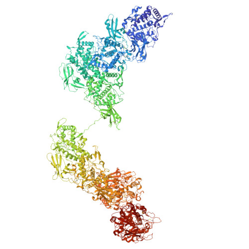
Cryo-EM structure of N-glycosylated Pomacea canaliculata hemocyanin


Experimental Data Snapshot

  • Method: ELECTRON MICROSCOPY
  • Resolution: 4.40 Å
  • Aggregation State: PARTICLE 
  • Reconstruction Method: SINGLE PARTICLE 

Starting Model: in silico
View more details

wwPDB Validation 3D Report Full Report

Validation slider image for 8TRZ

This is version 1.1 of the entry. See complete history

Literature

Cryo-EM structure of N-glycosylated Pomacea canaliculata hemocyanin provides insights into its role in the immune response of gastropods.

Brola, T.R.Chiumiento, I.R.Ituarte, S.Benitez, G.I.Gragera, M.Carazo, J.M.Heras, H.Otero, L.H.Dreon, M.S.

(2025) FEBS J 

  • DOI: https://doi.org/10.1111/febs.70378
  • Primary Citation Related Structures: 
    8TRZ

  • PubMed Abstract: 

    Hemocyanins are multifunctional soluble proteins found in most mollusks and some arthropods that may turn into a phenoloxidase-like enzyme (PO) associated with innate immune functions. To expand the structural understanding of gastropod hemocyanins and their physiological implications, here we report the single-particle cryogenic electron microscopy (cryo-EM) structure of Pomacea canaliculata snail hemocyanin (PcH) at 4.4 Å resolution along with its intrinsic and proteolytically induced PO activity. PcH shows a Megathura crenulata (giant keyhole limpet) hemocyanin (KLH)-type structure with a cylindrical shape, comprising 20 protomers assembled as di-pentamers of antiparallel asymmetric dimers, organized in a D5 symmetry. Each protomer comprises eight paralogous functional units (FUs) sharing conserved structural features typical of hemocyanins. The achieved map resolution allowed delineation of interaction networks among adjacent subunits across quaternary structure tiers-dimer, pentamer of dimers (decamer), and di-pentamer of dimers (di-decamer). Additionally, we identified six N-glycosylation sites per protomer, totaling 120 glycan trees in the overall structure. Kinetic analysis of intrinsic PO activity using catechol as substrate revealed a Michaelis constant (K M ) of 45.3 mm and a catalytic rate constant (k cat ) of 2.87 min -1 . This specific activity was enhanced by limited proteolysis using digestive and bacterial proteases. Potential protease cleavage sites were identified in silico, mapped onto the PcH model, and their accessibility assessed. Combined with molecular dynamics simulations, these findings suggest a structural basis for the PO induction mechanism. This study expands our knowledge of KLH-type hemocyanins and provides clues into their PO activation, which is triggered by endogenous and/or pathogen-associated proteases. This further underscores the role of molluscan hemocyanins in the innate immune system.


  • Organizational Affiliation
    • Instituto de Investigaciones Bioquímicas de La Plata "Prof. Dr. Rodolfo R. Brenner" (INIBIOLP), Consejo Nacional de Investigaciones Científicas y Técnicas (CONICET), Universidad Nacional de La Plata (UNLP), Argentina.

Macromolecule Content 

  • Total Structure Weight: 7,831.14 kDa 
  • Atom Count: 549,912 
  • Modeled Residue Count: 67,750 
  • Deposited Residue Count: 68,160 
  • Unique protein chains: 1

Macromolecules

Find similar proteins by:|  3D Structure
Entity ID: 1
MoleculeChains  Sequence LengthOrganismDetailsImage
Hemocyanin
A, B, C, D, E
A, B, C, D, E, F, G, H, I, J, K, L, M, N, O, P, Q, R, S, T
3,408Pomacea canaliculataMutation(s): 0 
UniProt
Find proteins for A0A2T7PFG7 (Pomacea canaliculata)
Explore A0A2T7PFG7 
Go to UniProtKB:  A0A2T7PFG7
Entity Groups
Sequence Clusters30% Identity50% Identity70% Identity90% Identity95% Identity100% Identity
UniProt GroupA0A2T7PFG7
Glycosylation
Glycosylation Sites: 6
Sequence Annotations
Expand
Reference Sequence

Oligosaccharides

Help  
Entity ID: 2
MoleculeChains Length2D Diagram GlycosylationD Interactions
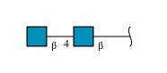
2-acetamido-2-deoxy-beta-D-glucopyranose-(1-4)-2-acetamido-2-deoxy-beta-D-glucopyranose
AB [auth 0],
AD [auth qA],
BC [auth RA],
CB [auth 2],
CC [auth SA],
AB [auth 0],
AD [auth qA],
BC [auth RA],
CB [auth 2],
CC [auth SA],
DA [auth d],
EA [auth e],
EC [auth UA],
FD [auth vA],
GA [auth g],
GC [auth WA],
GD [auth wA],
HB [auth 7],
IA [auth i],
IB [auth 8],
ID [auth yA],
KB [auth AA],
KD [auth 0A],
LC [auth bA],
MB [auth CA],
MC [auth cA],
NA [auth n],
OA [auth o],
OC [auth eA],
PD [auth 5A],
QA [auth q],
QC [auth gA],
RB [auth HA],
SA [auth s],
SB [auth IA],
U,
UB [auth KA],
VC [auth lA],
W,
WB [auth MA],
WC [auth mA],
XA [auth x],
Y,
YA [auth y],
YC [auth oA]
2N-Glycosylation
Glycosylation Resources
GlyTouCan: G42666HT
GlyCosmos: G42666HT
GlyGen: G42666HT
Entity ID: 3
MoleculeChains Length2D Diagram GlycosylationD Interactions
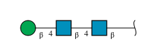
beta-D-mannopyranose-(1-4)-2-acetamido-2-deoxy-beta-D-glucopyranose-(1-4)-2-acetamido-2-deoxy-beta-D-glucopyranose
AA [auth a],
AC [auth QA],
BA [auth b],
BB [auth 1],
BD [auth rA],
AA [auth a],
AC [auth QA],
BA [auth b],
BB [auth 1],
BD [auth rA],
CA [auth c],
CD [auth sA],
DB [auth 3],
DC [auth TA],
DD [auth tA],
EB [auth 4],
ED [auth uA],
FA [auth f],
FB [auth 5],
FC [auth VA],
GB [auth 6],
HA [auth h],
HC [auth XA],
HD [auth xA],
IC [auth YA],
JA [auth j],
JB [auth 9],
JC [auth ZA],
JD [auth zA],
KA [auth k],
KC [auth aA],
LA [auth l],
LB [auth BA],
LD [auth 1A],
MA [auth m],
MD [auth 2A],
NB [auth DA],
NC [auth dA],
ND [auth 3A],
OB [auth EA],
OD [auth 4A],
PA [auth p],
PB [auth FA],
PC [auth fA],
QB [auth GA],
RA [auth r],
RC [auth hA],
SC [auth iA],
TA [auth t],
TB [auth JA],
TC [auth jA],
UA [auth u],
UC [auth kA],
V,
VA [auth v],
VB [auth LA],
WA [auth w],
X,
XB [auth NA],
XC [auth nA],
YB [auth OA],
Z,
ZA [auth z],
ZB [auth PA],
ZC [auth pA]
3N-Glycosylation
Glycosylation Resources
GlyTouCan: G15407YE
GlyCosmos: G15407YE
GlyGen: G15407YE

Small Molecules

Ligands 1 Unique
IDChains Name / Formula / InChI Key2D Diagram3D Interactions
NAG
(Subject of Investigation/LOI)

Query on NAG


Download:Ideal Coordinates CCD File
AE [auth K]
BE [auth L]
CE [auth M]
DE [auth N]
EE [auth O]
AE [auth K],
BE [auth L],
CE [auth M],
DE [auth N],
EE [auth O],
FE [auth P],
GE [auth Q],
HE [auth R],
IE [auth S],
JE [auth T],
QD [auth A],
RD [auth B],
SD [auth C],
TD [auth D],
UD [auth E],
VD [auth F],
WD [auth G],
XD [auth H],
YD [auth I],
ZD [auth J]
2-acetamido-2-deoxy-beta-D-glucopyranose
C8 H15 N O6
OVRNDRQMDRJTHS-FMDGEEDCSA-N

Experimental Data & Validation

Experimental Data

  • Method: ELECTRON MICROSCOPY
  • Resolution: 4.40 Å
  • Aggregation State: PARTICLE 
  • Reconstruction Method: SINGLE PARTICLE 
EM Software:
TaskSoftware PackageVersion
MODEL REFINEMENTPHENIX
RECONSTRUCTIONcryoSPARC
RECONSTRUCTIONScipion

Structure Validation

View Full Validation Report


Entry History 

& Funding Information

Deposition Data


Funding OrganizationLocationGrant Number
Agencia Nacional de Promocion Cientifica y Tecnologica (FONCYT)ArgentinaPICT-2021-00158
Agencia Nacional de Promocion Cientifica y Tecnologica (FONCYT)ArgentinaPICT-2020- SERIEA-03047

Revision History  (Full details and data files)

  • Version 1.0: 2025-04-02
    Type: Initial release
  • Version 1.1: 2026-04-15
    Changes: Data collection, Database references