8RLN

Crystal structure of human adenosine A2A receptor (construct A2A-PSB2-bRIL) complexed with the partial antagonist LUF5834 at the orthosteric pocket


Experimental Data Snapshot

  • Method: X-RAY DIFFRACTION
  • Resolution: 2.43 Å
  • R-Value Free: 0.281 
  • R-Value Work: 0.203 
  • R-Value Observed: 0.207 

wwPDB Validation   3D Report Full Report


Ligand Structure Quality Assessment 


This is version 1.0 of the entry. See complete history


Literature

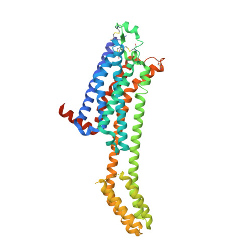
Structural Insights into Partial Activation of the Prototypic G Protein-Coupled Adenosine A 2A Receptor.

Claff, T.Mahardhika, A.B.Vaassen, V.J.Schlegel, J.G.Vielmuth, C.Weisse, R.H.Strater, N.Muller, C.E.

(2024) Acs Pharmacol Transl Sci 7: 1415-1425

  • DOI: https://doi.org/10.1021/acsptsci.4c00051
  • Primary Citation of Related Structures:  
    8RLN

  • PubMed Abstract: 

    The adenosine A 2A receptor (A 2A AR) belongs to the rhodopsin-like G protein-coupled receptor (GPCR) family, which constitutes the largest class of GPCRs. Partial agonists show reduced efficacy as compared to physiological agonists and can even act as antagonists in the presence of a full agonist. Here, we determined an X-ray crystal structure of the partial A 2A AR agonist 2-amino-6-[(1 H -imidazol-2-ylmethyl)sulfanyl]-4- p -hydroxyphenyl-3,5-pyridinedicarbonitrile (LUF5834) in complex with the A 2A AR construct A 2A -PSB2-bRIL, stabilized in its inactive conformation and being devoid of any mutations in the ligand binding pocket. The determined high-resolution structure (2.43 Å) resolved water networks and crucial binding pocket interactions. A direct hydrogen bond of the p -hydroxy group of LUF5834 with T88 3.36 was observed, an amino acid that was mutated to alanine in the most frequently used A 2A AR crystallization constructs thus preventing the discovery of its interactions in most of the previous A 2A AR co-crystal structures. G protein dissociation studies confirmed partial agonistic activity of LUF5834 as compared to that of the full agonist N -ethylcarboxamidoadenosine (NECA). In contrast to NECA, the partial agonist was still able to bind to the receptor construct locked in its inactive conformation by an S91 3.39 K mutation, although with an affinity lower than that at the native receptor. This could explain the compound's partial agonistic activity: while full A 2A AR agonists bind exclusively to the active conformation, likely following conformational selection, partial agonists bind to active as well as inactive conformations, showing higher affinity for the active conformation. This might be a general mechanism of partial agonism also applicable to other GPCRs.


  • Organizational Affiliation

    PharmaCenter Bonn & Pharmaceutical Institute, Department of Pharmaceutical & Medicinal Chemistry, University of Bonn, Bonn 53113, Germany.


Macromolecules
Find similar proteins by:  (by identity cutoff)  |  3D Structure
Entity ID: 1
MoleculeChains Sequence LengthOrganismDetailsImage
Adenosine receptor A2a,Soluble cytochrome b562431Homo sapiensMutation(s): 0 
Gene Names: ADORA2AADORA2cybC
UniProt & NIH Common Fund Data Resources
Find proteins for P0ABE7 (Escherichia coli)
Explore P0ABE7 
Go to UniProtKB:  P0ABE7
Find proteins for P29274 (Homo sapiens)
Explore P29274 
Go to UniProtKB:  P29274
PHAROS:  P29274
GTEx:  ENSG00000128271 
Entity Groups  
Sequence Clusters30% Identity50% Identity70% Identity90% Identity95% Identity100% Identity
UniProt GroupsP0ABE7P29274
Sequence Annotations
Expand
  • Reference Sequence
Small Molecules
Ligands 6 Unique
IDChains Name / Formula / InChI Key2D Diagram3D Interactions
CLR
Query on CLR

Download Ideal Coordinates CCD File 
B [auth A],
C [auth A],
D [auth A]
CHOLESTEROL
C27 H46 O
HVYWMOMLDIMFJA-DPAQBDIFSA-N
OLB
Query on OLB

Download Ideal Coordinates CCD File 
I [auth A](2S)-2,3-dihydroxypropyl (9Z)-octadec-9-enoate
C21 H40 O4
RZRNAYUHWVFMIP-QJRAZLAKSA-N
OLC
Query on OLC

Download Ideal Coordinates CCD File 
H [auth A],
J [auth A],
N [auth A]
(2R)-2,3-dihydroxypropyl (9Z)-octadec-9-enoate
C21 H40 O4
RZRNAYUHWVFMIP-GDCKJWNLSA-N
A1H1S (Subject of Investigation/LOI)
Query on A1H1S

Download Ideal Coordinates CCD File 
L [auth A]2-azanyl-4-(4-hydroxyphenyl)-6-(1~{H}-imidazol-2-ylmethylsulfanyl)pyridine-3,5-dicarbonitrile
C17 H12 N6 O S
OFHKDLYFKPBXER-UHFFFAOYSA-N
OLA
Query on OLA

Download Ideal Coordinates CCD File 
E [auth A],
F [auth A],
G [auth A],
M [auth A]
OLEIC ACID
C18 H34 O2
ZQPPMHVWECSIRJ-KTKRTIGZSA-N
PEG
Query on PEG

Download Ideal Coordinates CCD File 
K [auth A]DI(HYDROXYETHYL)ETHER
C4 H10 O3
MTHSVFCYNBDYFN-UHFFFAOYSA-N
Experimental Data & Validation

Experimental Data

  • Method: X-RAY DIFFRACTION
  • Resolution: 2.43 Å
  • R-Value Free: 0.281 
  • R-Value Work: 0.203 
  • R-Value Observed: 0.207 
  • Space Group: C 2 2 21
Unit Cell:
Length ( Å )Angle ( ˚ )
a = 39.556α = 90
b = 179.693β = 90
c = 140.454γ = 90
Software Package:
Software NamePurpose
PHENIXrefinement
XDSdata reduction
Aimlessdata scaling
STARANISOdata scaling
autoPROCdata processing

Structure Validation

View Full Validation Report


Ligand Structure Quality Assessment 


Entry History & Funding Information

Deposition Data


Funding OrganizationLocationGrant Number
German Research Foundation (DFG)GermanyGRK 1873
German Research Foundation (DFG)GermanyGRK 2873
German Research Foundation (DFG)GermanySFB 1328
German Research Foundation (DFG)GermanySFB 1423, project number 421152132, subproject A6
German Federal Ministry for Education and ResearchGermanyBonn Graduate School of Drug Sciences (BIGS-DrugS)

Revision History  (Full details and data files)

  • Version 1.0: 2024-06-19
    Type: Initial release