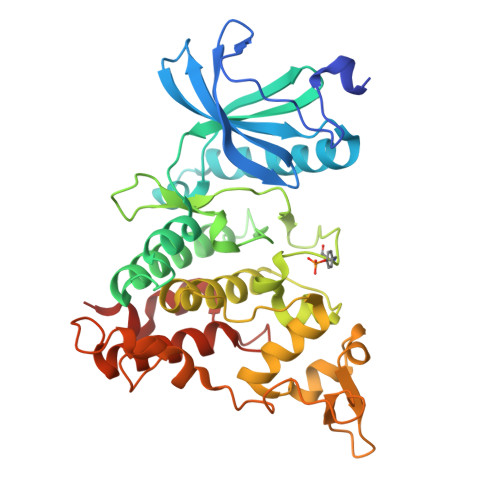
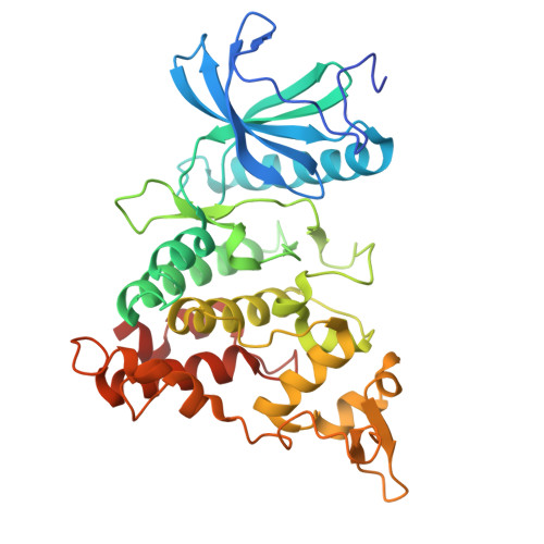
8R8E

DYRK1a in Complex with 2-Cyclopentyl-7-iodo-1H-indole-3-carbonitrile


Experimental Data Snapshot

  • Method: X-RAY DIFFRACTION
  • Resolution: 2.22 Å
  • R-Value Free: 0.234 
  • R-Value Work: 0.199 
  • R-Value Observed: 0.200 

Starting Model: experimental
View more details

wwPDB Validation   3D Report Full Report


Ligand Structure Quality Assessment 


This is version 1.1 of the entry. See complete history


Literature

Halogen Bonding on Water─A Drop in the Ocean?

Engelhardt, M.U.Zimmermann, M.O.Dammann, M.Stahlecker, J.Poso, A.Kronenberger, T.Kunick, C.Stehle, T.Boeckler, F.M.

(2024) J Chem Theory Comput 

  • DOI: https://doi.org/10.1021/acs.jctc.4c00834
  • Primary Citation of Related Structures:  
    8R8E

  • PubMed Abstract: 

    Halogen bonding is a valuable interaction in drug design, offering an unconventional way to influence affinity and selectivity by leveraging the halogen atoms' ability to form directional bonds. The present study evaluates halogen-water interactions within protein binding sites, demonstrating that targeting a water molecule via halogen bonding can in specific cases contribute beneficially to ligand binding. In solving and examining the crystal structure of 2-cyclopentyl-7-iodo-1 H -indole-3-carbonitrile bound to DYRK1a kinase, we identified a notable iodine-water interaction, where water accepts a halogen bond with good geometric and energetic features. This starting point triggered further investigations into the prevalence of such interactions across various halogen-bearing ligands (chlorine, bromine, iodine) in the PDB. Using QM calculations (MP2/TZVPP), we highlight the versatility and potential benefits of such halogen-water interactions, particularly when the water molecule is a stable part of the binding site's structured environment. While the interaction energies with water are lower compared to other typical halogen bond acceptors, we deem this different binding strength essential for reducing desolvation costs. We suggest that "interstitial" water molecules, as stable parts of the binding site engaging in multiple strong interactions, could be prime targets for halogen bonding. Further systematic studies, combining high-resolution crystal structures and quantum chemistry, are required to scrutinize whether halogen bonding on water is more than a "drop in the ocean".


  • Organizational Affiliation

    Laboratory for Molecular Design & Pharmaceutical Biophysics, Institute of Pharmaceutical Sciences, Department of Pharmacy and Biochemistry, Eberhard Karls Universität Tübingen, 72076 Tübingen, Germany.


Macromolecules
Find similar proteins by:  (by identity cutoff)  |  3D Structure
Entity ID: 1
MoleculeChains Sequence LengthOrganismDetailsImage
Dual specificity tyrosine-phosphorylation-regulated kinase 1A
A, B, C
361Homo sapiensMutation(s): 0 
Gene Names: DYRK1ADYRKMNBMNBH
EC: 2.7.12.1 (PDB Primary Data), 2.7.11.23 (UniProt)
UniProt & NIH Common Fund Data Resources
Find proteins for Q13627 (Homo sapiens)
Explore Q13627 
Go to UniProtKB:  Q13627
PHAROS:  Q13627
GTEx:  ENSG00000157540 
Entity Groups  
Sequence Clusters30% Identity50% Identity70% Identity90% Identity95% Identity100% Identity
UniProt GroupQ13627
Sequence Annotations
Expand
  • Reference Sequence
Find similar proteins by:  (by identity cutoff)  |  3D Structure
Entity ID: 2
MoleculeChains Sequence LengthOrganismDetailsImage
Dual specificity tyrosine-phosphorylation-regulated kinase 1A361Homo sapiensMutation(s): 0 
Gene Names: DYRK1A
EC: 2.7.12.1 (UniProt), 2.7.11.23 (UniProt)
UniProt & NIH Common Fund Data Resources
Find proteins for Q13627 (Homo sapiens)
Explore Q13627 
Go to UniProtKB:  Q13627
PHAROS:  Q13627
GTEx:  ENSG00000157540 
Entity Groups  
Sequence Clusters30% Identity50% Identity70% Identity90% Identity95% Identity100% Identity
UniProt GroupQ13627
Sequence Annotations
Expand
  • Reference Sequence
Small Molecules
Ligands 9 Unique
IDChains Name / Formula / InChI Key2D Diagram3D Interactions
YOX (Subject of Investigation/LOI)
Query on YOX

Download Ideal Coordinates CCD File 
AB [auth D],
E [auth A],
PA [auth C],
Z [auth B]
2-cyclopentyl-7-iodanyl-1~{H}-indole-3-carbonitrile
C14 H13 I N2
BBYDGWHLEKHRAJ-UHFFFAOYSA-N
EPE
Query on EPE

Download Ideal Coordinates CCD File 
AA [auth B],
CA [auth B],
H [auth A],
RA [auth C]
4-(2-HYDROXYETHYL)-1-PIPERAZINE ETHANESULFONIC ACID
C8 H18 N2 O4 S
JKMHFZQWWAIEOD-UHFFFAOYSA-N
1PE
Query on 1PE

Download Ideal Coordinates CCD File 
CB [auth D],
F [auth A],
I [auth A],
QA [auth C],
Y [auth B]
PENTAETHYLENE GLYCOL
C10 H22 O6
JLFNLZLINWHATN-UHFFFAOYSA-N
PG4
Query on PG4

Download Ideal Coordinates CCD File 
G [auth A]TETRAETHYLENE GLYCOL
C8 H18 O5
UWHCKJMYHZGTIT-UHFFFAOYSA-N
PEG
Query on PEG

Download Ideal Coordinates CCD File 
HA [auth B],
J [auth A],
P [auth A]
DI(HYDROXYETHYL)ETHER
C4 H10 O3
MTHSVFCYNBDYFN-UHFFFAOYSA-N
SO4
Query on SO4

Download Ideal Coordinates CCD File 
EB [auth D]
FB [auth D]
GB [auth D]
HB [auth D]
IB [auth D]
EB [auth D],
FB [auth D],
GB [auth D],
HB [auth D],
IB [auth D],
JA [auth B],
JB [auth D],
KA [auth B],
KB [auth D],
LA [auth B],
MA [auth B],
NA [auth B],
Q [auth A],
R [auth A],
S [auth A],
T [auth A],
U [auth A],
V [auth A],
VA [auth C],
W [auth A],
WA [auth C],
X [auth A],
XA [auth C],
YA [auth C],
ZA [auth C]
SULFATE ION
O4 S
QAOWNCQODCNURD-UHFFFAOYSA-L
PO4
Query on PO4

Download Ideal Coordinates CCD File 
LB [auth D]PHOSPHATE ION
O4 P
NBIIXXVUZAFLBC-UHFFFAOYSA-K
GOL
Query on GOL

Download Ideal Coordinates CCD File 
BA [auth B]
BB [auth D]
DB [auth D]
EA [auth B]
GA [auth B]
BA [auth B],
BB [auth D],
DB [auth D],
EA [auth B],
GA [auth B],
K [auth A],
L [auth A],
O [auth A],
OA [auth C],
SA [auth C],
TA [auth C]
GLYCEROL
C3 H8 O3
PEDCQBHIVMGVHV-UHFFFAOYSA-N
EDO
Query on EDO

Download Ideal Coordinates CCD File 
DA [auth B]
FA [auth B]
IA [auth B]
M [auth A]
N [auth A]
DA [auth B],
FA [auth B],
IA [auth B],
M [auth A],
N [auth A],
UA [auth C]
1,2-ETHANEDIOL
C2 H6 O2
LYCAIKOWRPUZTN-UHFFFAOYSA-N
Modified Residues  1 Unique
IDChains TypeFormula2D DiagramParent
PTR
Query on PTR
A, B, C
L-PEPTIDE LINKINGC9 H12 N O6 PTYR
Experimental Data & Validation

Experimental Data

  • Method: X-RAY DIFFRACTION
  • Resolution: 2.22 Å
  • R-Value Free: 0.234 
  • R-Value Work: 0.199 
  • R-Value Observed: 0.200 
  • Space Group: C 1 2 1
Unit Cell:
Length ( Å )Angle ( ˚ )
a = 244.236α = 90
b = 64.473β = 115.58
c = 147.332γ = 90
Software Package:
Software NamePurpose
PHENIXrefinement
XDSdata reduction
XDSdata scaling
PHASERphasing

Structure Validation

View Full Validation Report


Ligand Structure Quality Assessment 


Entry History & Funding Information

Deposition Data


Funding OrganizationLocationGrant Number
Not funded--

Revision History  (Full details and data files)

  • Version 1.0: 2024-09-25
    Type: Initial release
  • Version 1.1: 2024-10-16
    Changes: Structure summary