Halogen Bonding on Water─A Drop in the Ocean?
Engelhardt, M.U., Zimmermann, M.O., Dammann, M., Stahlecker, J., Poso, A., Kronenberger, T., Kunick, C., Stehle, T., Boeckler, F.M.(2024) J Chem Theory Comput 20: 10716-10730
- PubMed: 39291905 Search on PubMed
- DOI: https://doi.org/10.1021/acs.jctc.4c00834
- Primary Citation Related Structures:
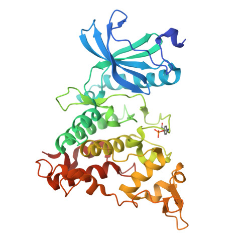
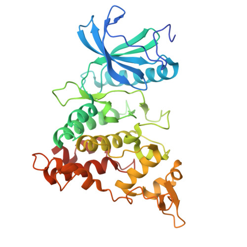
8R8E - PubMed Abstract:
Halogen bonding is a valuable interaction in drug design, offering an unconventional way to influence affinity and selectivity by leveraging the halogen atoms' ability to form directional bonds. The present study evaluates halogen-water interactions within protein binding sites, demonstrating that targeting a water molecule via halogen bonding can in specific cases contribute beneficially to ligand binding. In solving and examining the crystal structure of 2-cyclopentyl-7-iodo-1 H -indole-3-carbonitrile bound to DYRK1a kinase, we identified a notable iodine-water interaction, where water accepts a halogen bond with good geometric and energetic features. This starting point triggered further investigations into the prevalence of such interactions across various halogen-bearing ligands (chlorine, bromine, iodine) in the PDB. Using QM calculations (MP2/TZVPP), we highlight the versatility and potential benefits of such halogen-water interactions, particularly when the water molecule is a stable part of the binding site's structured environment. While the interaction energies with water are lower compared to other typical halogen bond acceptors, we deem this different binding strength essential for reducing desolvation costs. We suggest that "interstitial" water molecules, as stable parts of the binding site engaging in multiple strong interactions, could be prime targets for halogen bonding. Further systematic studies, combining high-resolution crystal structures and quantum chemistry, are required to scrutinize whether halogen bonding on water is more than a "drop in the ocean".
- Laboratory for Molecular Design & Pharmaceutical Biophysics, Institute of Pharmaceutical Sciences, Department of Pharmacy and Biochemistry, Eberhard Karls Universität Tübingen, 72076 Tübingen, Germany.
Organizational Affiliation: