8OUO

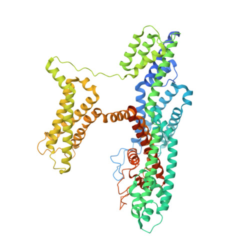
Human TPC2 in Complex with Antagonist (S)-SG-094


Experimental Data Snapshot

  • Method: ELECTRON MICROSCOPY
  • Resolution: 3.00 Å
  • Aggregation State: PARTICLE 
  • Reconstruction Method: SINGLE PARTICLE 

wwPDB Validation   3D Report Full Report


This is version 1.1 of the entry. See complete history


Literature

Structural basis for inhibition of the lysosomal two-pore channel TPC2 by a small molecule antagonist.

Chi, G.Jaslan, D.Kudrina, V.Bock, J.Li, H.Pike, A.C.W.Rautenberg, S.Krogsaeter, E.Bohstedt, T.Wang, D.McKinley, G.Fernandez-Cid, A.Mukhopadhyay, S.M.M.Burgess-Brown, N.A.Keller, M.Bracher, F.Grimm, C.Durr, K.L.

(2024) Structure 32: 1137-1149.e4

  • DOI: https://doi.org/10.1016/j.str.2024.05.005
  • Primary Citation of Related Structures:  
    8OUO

  • PubMed Abstract: 

    Two pore channels are lysosomal cation channels with crucial roles in tumor angiogenesis and viral release from endosomes. Inhibition of the two-pore channel 2 (TPC2) has emerged as potential therapeutic strategy for the treatment of cancers and viral infections, including Ebola and COVID-19. Here, we demonstrate that antagonist SG-094, a synthetic analog of the Chinese alkaloid medicine tetrandrine with increased potency and reduced toxicity, induces asymmetrical structural changes leading to a single binding pocket at only one intersubunit interface within the asymmetrical dimer. Supported by functional characterization of mutants by Ca 2+ imaging and patch clamp experiments, we identify key residues in S1 and S4 involved in compound binding to the voltage sensing domain II. SG-094 arrests IIS4 in a downward shifted state which prevents pore opening via the IIS4/S5 linker, hence resembling gating modifiers of canonical VGICs. These findings may guide the rational development of new therapeutics antagonizing TPC2 activity.


  • Organizational Affiliation

    Centre for Medicines Discovery, Nuffield Department of Medicine, University of Oxford, Nuffield Department of Medicine Research Building, Oxford OX3 7FZ, UK; Structural Genomics Consortium, Nuffield Department of Medicine, University of Oxford, Nuffield Department of Medicine Research Building, Oxford OX3 7FZ, UK. Electronic address: gamma.chi@cmd.ox.ac.uk.


Macromolecules
Find similar proteins by:  (by identity cutoff)  |  3D Structure
Entity ID: 1
MoleculeChains Sequence LengthOrganismDetailsImage
Two pore channel protein 2
A, B
752Homo sapiensMutation(s): 2 
Gene Names: TPCN2TPC2
Membrane Entity: Yes 
UniProt & NIH Common Fund Data Resources
Find proteins for Q8NHX9 (Homo sapiens)
Explore Q8NHX9 
Go to UniProtKB:  Q8NHX9
PHAROS:  Q8NHX9
GTEx:  ENSG00000162341 
Entity Groups  
Sequence Clusters30% Identity50% Identity70% Identity90% Identity95% Identity100% Identity
UniProt GroupQ8NHX9
Sequence Annotations
Expand
  • Reference Sequence
Small Molecules
Ligands 5 Unique
IDChains Name / Formula / InChI Key2D Diagram3D Interactions
Q7G
Query on Q7G

Download Ideal Coordinates CCD File 
D [auth A],
J [auth A],
P [auth B]
2-{[(4-O-alpha-D-glucopyranosyl-alpha-D-glucopyranosyl)oxy]methyl}-4-{[(3beta,9beta,14beta,17beta,25R)-spirost-5-en-3-yl]oxy}butyl 4-O-alpha-D-glucopyranosyl-alpha-D-glucopyranoside
C56 H92 O25
LKBFXDNKNZXHHW-NXLTVWPKSA-N
PLD
Query on PLD

Download Ideal Coordinates CCD File 
C [auth A],
K [auth B]
di-heneicosanoyl phosphatidyl choline
C50 H101 N O8 P
QFFSGJSMHPWZOB-QSCHNALKSA-O
PCF
Query on PCF

Download Ideal Coordinates CCD File 
F [auth A],
L [auth B]
1,2-DIACYL-SN-GLYCERO-3-PHOSHOCHOLINE
C40 H80 N O8 P
KILNVBDSWZSGLL-KXQOOQHDSA-N
Y01
Query on Y01

Download Ideal Coordinates CCD File 
G [auth A]
H [auth A]
I [auth A]
M [auth B]
N [auth B]
G [auth A],
H [auth A],
I [auth A],
M [auth B],
N [auth B],
O [auth B]
CHOLESTEROL HEMISUCCINATE
C31 H50 O4
WLNARFZDISHUGS-MIXBDBMTSA-N
W4E (Subject of Investigation/LOI)
Query on W4E

Download Ideal Coordinates CCD File 
E [auth A](1S)-6-methoxy-2-methyl-7-phenoxy-1-[(4-phenoxyphenyl)methyl]-3,4-dihydro-1H-isoquinoline
C30 H29 N O3
BTNHPUSFPKFAPU-NDEPHWFRSA-N
Experimental Data & Validation

Experimental Data

  • Method: ELECTRON MICROSCOPY
  • Resolution: 3.00 Å
  • Aggregation State: PARTICLE 
  • Reconstruction Method: SINGLE PARTICLE 

Structure Validation

View Full Validation Report


Entry History & Funding Information

Deposition Data


Funding OrganizationLocationGrant Number
Wellcome TrustUnited Kingdom--

Revision History  (Full details and data files)

  • Version 1.0: 2024-06-12
    Type: Initial release
  • Version 1.1: 2024-08-21
    Changes: Data collection, Database references