8KBZ

Cryo-EM structure of human ATG9A in LMNG micelles

  • Classification: MEMBRANE PROTEIN
  • Organism(s): Homo sapiens
  • Expression System: Homo sapiens
  • Mutation(s): No 

  • Deposited: 2023-08-04 Released: 2024-08-07 
  • Deposition Author(s): Yang, W., Goran, S.
  • Funding Organization(s): National Natural Science Foundation of China (NSFC), Ministry of Science and Technology (MoST, China)

Experimental Data Snapshot

  • Method: ELECTRON MICROSCOPY
  • Resolution: 3.97 Å
  • Aggregation State: PARTICLE 
  • Reconstruction Method: SINGLE PARTICLE 

wwPDB Validation   3D Report Full Report


This is version 1.1 of the entry. See complete history


Literature

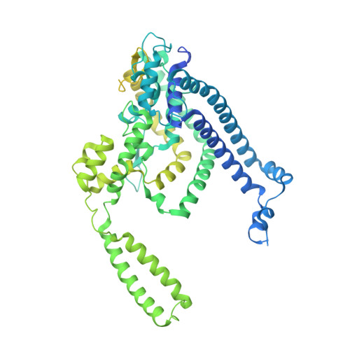
Structural basis for lipid transfer by the ATG2A-ATG9A complex.

Wang, Y.Dahmane, S.Ti, R.Mai, X.Zhu, L.Carlson, L.A.Stjepanovic, G.

(2024) Nat Struct Mol Biol 

  • DOI: https://doi.org/10.1038/s41594-024-01376-6
  • Primary Citation of Related Structures:  
    8KBX, 8KBY, 8KBZ, 8KC3, 8Y1L

  • PubMed Abstract: 

    Autophagy is characterized by the formation of double-membrane vesicles called autophagosomes. Autophagy-related proteins (ATGs) 2A and 9A have an essential role in autophagy by mediating lipid transfer and re-equilibration between membranes for autophagosome formation. Here we report the cryo-electron microscopy structures of human ATG2A in complex with WD-repeat protein interacting with phosphoinositides 4 (WIPI4) at 3.2 Å and the ATG2A-WIPI4-ATG9A complex at 7 Å global resolution. On the basis of molecular dynamics simulations, we propose a mechanism of lipid extraction from the donor membranes. Our analysis revealed 3:1 stoichiometry of the ATG9A-ATG2A complex, directly aligning the ATG9A lateral pore with ATG2A lipid transfer cavity, and an interaction of the ATG9A trimer with both the N-terminal and the C-terminal tip of rod-shaped ATG2A. Cryo-electron tomography of ATG2A liposome-binding states showed that ATG2A tethers lipid vesicles at different orientations. In summary, this study provides a molecular basis for the growth of the phagophore membrane and lends structural insights into spatially coupled lipid transport and re-equilibration during autophagosome formation.


  • Organizational Affiliation

    Kobilka Institute of Innovative Drug Discovery, School of Medicine, The Chinese University of Hong Kong, Shenzhen, Shenzhen, China.


Macromolecules
Find similar proteins by:  (by identity cutoff)  |  3D Structure
Entity ID: 1
MoleculeChains Sequence LengthOrganismDetailsImage
Autophagy-related protein 9A
A, B, C
839Homo sapiensMutation(s): 0 
Gene Names: ATG9AAPG9L1
UniProt & NIH Common Fund Data Resources
Find proteins for Q7Z3C6 (Homo sapiens)
Explore Q7Z3C6 
Go to UniProtKB:  Q7Z3C6
PHAROS:  Q7Z3C6
GTEx:  ENSG00000198925 
Entity Groups  
Sequence Clusters30% Identity50% Identity70% Identity90% Identity95% Identity100% Identity
UniProt GroupQ7Z3C6
Sequence Annotations
Expand
  • Reference Sequence
Experimental Data & Validation

Experimental Data

  • Method: ELECTRON MICROSCOPY
  • Resolution: 3.97 Å
  • Aggregation State: PARTICLE 
  • Reconstruction Method: SINGLE PARTICLE 

Structure Validation

View Full Validation Report


Entry History & Funding Information

Deposition Data


Funding OrganizationLocationGrant Number
National Natural Science Foundation of China (NSFC)China31950410540
Ministry of Science and Technology (MoST, China)ChinaQN2021032004L

Revision History  (Full details and data files)

  • Version 1.0: 2024-08-07
    Type: Initial release
  • Version 1.1: 2024-09-04
    Changes: Data collection, Database references