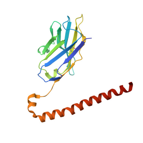
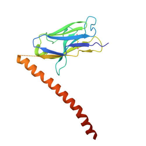
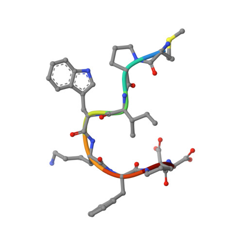
8JYQ

Crystal structure of cancer-specific anti-HER2 antibody H2Mab-214 in complex with epitope peptide


Experimental Data Snapshot

  • Method: X-RAY DIFFRACTION
  • Resolution: 1.75 Å
  • R-Value Free: 0.233 
  • R-Value Work: 0.196 
  • R-Value Observed: 0.198 

wwPDB Validation   3D Report Full Report


This is version 1.2 of the entry. See complete history


Literature

Locally misfolded HER2 expressed on cancer cells is a promising target for development of cancer-specific antibodies.

Arimori, T.Mihara, E.Suzuki, H.Ohishi, T.Tanaka, T.Kaneko, M.K.Takagi, J.Kato, Y.

(2024) Structure 32: 536-549.e5

  • DOI: https://doi.org/10.1016/j.str.2024.02.007
  • Primary Citation of Related Structures:  
    8JYQ, 8JYR

  • PubMed Abstract: 

    Overexpression of human epidermal growth factor receptor 2 (HER2) in breast and gastric cancers is associated with a poor prognosis, making it an important therapeutic target. Here, we establish a novel cancer-specific anti-HER2 antibody, H 2 Mab-214. H 2 Mab-214 reacts with HER2 on cancer cells, but unlike the therapeutic antibody trastuzumab, it does not react with HER2 on normal cells in flow cytometry measurements. A crystal structure suggests that H 2 Mab-214 recognizes a structurally disrupted region in the HER2 domain IV, which normally forms a β-sheet. We show that this misfolding is inducible by site-directed mutagenesis mimicking the disulfide bond defects that also may occur in cancer cells, indicating that the local misfolding in the Cys-rich domain IV governs the cancer-specificity of H 2 Mab-214. Furthermore, we show that H 2 Mab-214 effectively suppresses tumor growth in xenograft mouse models. Our findings offer a potential strategy for developing cancer-specific therapeutic antibodies that target partially misfolded cell surface receptors.


  • Organizational Affiliation

    Institute for Protein Research, Osaka University, 3-2. Yamadaoka, Suita, Osaka 565-0871, Japan. Electronic address: arimori@protein.osaka-u.ac.jp.


Macromolecules
Find similar proteins by:  (by identity cutoff)  |  3D Structure
Entity ID: 1
MoleculeChains Sequence LengthOrganismDetailsImage
H2CasMab-1 VH(S112C),SARAH
A, D
174Mus musculusHomo sapiens
This entity is chimeric
Mutation(s): 0 
Entity Groups  
Sequence Clusters30% Identity50% Identity70% Identity90% Identity95% Identity100% Identity
Sequence Annotations
Expand
  • Reference Sequence
Find similar proteins by:  (by identity cutoff)  |  3D Structure
Entity ID: 2
MoleculeChains Sequence LengthOrganismDetailsImage
H2CasMab-1 VL,SARAH(S37C)
B, E
170Mus musculusHomo sapiens
This entity is chimeric
Mutation(s): 0 
Entity Groups  
Sequence Clusters30% Identity50% Identity70% Identity90% Identity95% Identity100% Identity
Sequence Annotations
Expand
  • Reference Sequence

Find similar proteins by:  Sequence   |   3D Structure  

Entity ID: 3
MoleculeChains Sequence LengthOrganismDetailsImage
H2CasMab-1 epitope peptide
C, F
8Homo sapiensMutation(s): 0 
Sequence Annotations
Expand
  • Reference Sequence
Experimental Data & Validation

Experimental Data

  • Method: X-RAY DIFFRACTION
  • Resolution: 1.75 Å
  • R-Value Free: 0.233 
  • R-Value Work: 0.196 
  • R-Value Observed: 0.198 
  • Space Group: P 1 21 1
Unit Cell:
Length ( Å )Angle ( ˚ )
a = 73.143α = 90
b = 62.793β = 94.419
c = 78.122γ = 90
Software Package:
Software NamePurpose
PHENIXrefinement
XDSdata reduction
XDSdata scaling
PHASERphasing

Structure Validation

View Full Validation Report


Entry History & Funding Information

Deposition Data


Funding OrganizationLocationGrant Number
Japan Society for the Promotion of Science (JSPS)Japan21H02416
Japan Agency for Medical Research and Development (AMED)Japan20am0101075

Revision History  (Full details and data files)

  • Version 1.0: 2024-03-13
    Type: Initial release
  • Version 1.1: 2024-03-27
    Changes: Database references
  • Version 1.2: 2024-05-15
    Changes: Database references