8JYQ | pdb_00008jyq

Crystal structure of cancer-specific anti-HER2 antibody H2Mab-214 in complex with epitope peptide


Experimental Data Snapshot

  • Method: X-RAY DIFFRACTION
  • Resolution: 1.75 Å
  • R-Value Free: 
    0.233 (Depositor), 0.266 (DCC) 
  • R-Value Work: 
    0.196 (Depositor) 
  • R-Value Observed: 
    0.198 (Depositor) 

Starting Models: experimental
View more details

wwPDB Validation 3D Report Full Report

Validation slider image for 8JYQ

This is version 1.3 of the entry. See complete history

Literature

Locally misfolded HER2 expressed on cancer cells is a promising target for development of cancer-specific antibodies.

Arimori, T.Mihara, E.Suzuki, H.Ohishi, T.Tanaka, T.Kaneko, M.K.Takagi, J.Kato, Y.

(2024) Structure 32: 536-549.e5

  • DOI: https://doi.org/10.1016/j.str.2024.02.007
  • Primary Citation Related Structures: 
    8JYQ, 8JYR

  • PubMed Abstract: 

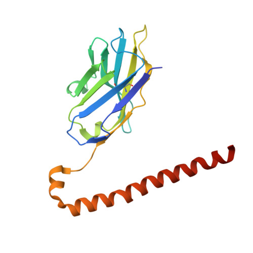
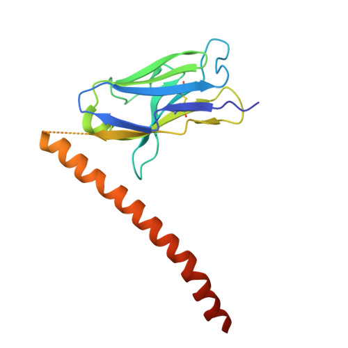
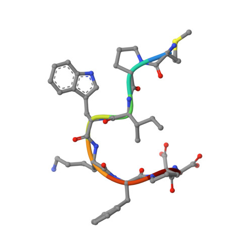
    Overexpression of human epidermal growth factor receptor 2 (HER2) in breast and gastric cancers is associated with a poor prognosis, making it an important therapeutic target. Here, we establish a novel cancer-specific anti-HER2 antibody, H 2 Mab-214. H 2 Mab-214 reacts with HER2 on cancer cells, but unlike the therapeutic antibody trastuzumab, it does not react with HER2 on normal cells in flow cytometry measurements. A crystal structure suggests that H 2 Mab-214 recognizes a structurally disrupted region in the HER2 domain IV, which normally forms a β-sheet. We show that this misfolding is inducible by site-directed mutagenesis mimicking the disulfide bond defects that also may occur in cancer cells, indicating that the local misfolding in the Cys-rich domain IV governs the cancer-specificity of H 2 Mab-214. Furthermore, we show that H 2 Mab-214 effectively suppresses tumor growth in xenograft mouse models. Our findings offer a potential strategy for developing cancer-specific therapeutic antibodies that target partially misfolded cell surface receptors.


  • Organizational Affiliation
    • Institute for Protein Research, Osaka University, 3-2. Yamadaoka, Suita, Osaka 565-0871, Japan. Electronic address: arimori@protein.osaka-u.ac.jp.

Macromolecule Content 

  • Total Structure Weight: 79.47 kDa 
  • Atom Count: 5,592 
  • Modeled Residue Count: 673 
  • Deposited Residue Count: 704 
  • Unique protein chains: 3

Macromolecules

Find similar proteins by:|  3D Structure
Entity ID: 1
MoleculeChains  Sequence LengthOrganismDetailsImage
H2CasMab-1 VH(S112C),SARAH
A, D
174Mus musculusHomo sapiens
This entity is chimeric
Mutation(s): 0 
Find similar proteins by:|  3D Structure
Entity ID: 2
MoleculeChains  Sequence LengthOrganismDetailsImage
H2CasMab-1 VL,SARAH(S37C)
B, E
170Mus musculusHomo sapiens
This entity is chimeric
Mutation(s): 0 
Find similar proteins by:  Sequence   |   3D Structure  
Entity ID: 3
MoleculeChains  Sequence LengthOrganismDetailsImage
H2CasMab-1 epitope peptide
C, F
8Homo sapiensMutation(s): 0 
EC: 2.7.10.1
UniProt & NIH Common Fund Data Resources
Find proteins for P04626 (Homo sapiens)
Explore P04626 
Go to UniProtKB:  P04626
PHAROS:  P04626
GTEx:  ENSG00000141736 
Entity Groups
UniProt GroupP04626
Sequence Annotations
Expand
Reference Sequence

Experimental Data & Validation

Experimental Data

  • Method: X-RAY DIFFRACTION
  • Resolution: 1.75 Å
  • R-Value Free:  0.233 (Depositor), 0.266 (DCC) 
  • R-Value Work:  0.196 (Depositor) 
  • R-Value Observed: 0.198 (Depositor) 
Space Group: P 1 21 1
Unit Cell:
Length ( Å )Angle ( ˚ )
a = 73.143α = 90
b = 62.793β = 94.419
c = 78.122γ = 90
Software Package:
Software NamePurpose
PHENIXrefinement
XDSdata reduction
XDSdata scaling
PHASERphasing

Structure Validation

View Full Validation Report


Entry History 

& Funding Information

Deposition Data


Funding OrganizationLocationGrant Number
Japan Society for the Promotion of Science (JSPS)Japan21H02416
Japan Agency for Medical Research and Development (AMED)Japan20am0101075

Revision History  (Full details and data files)

  • Version 1.0: 2024-03-13
    Type: Initial release
  • Version 1.1: 2024-03-27
    Changes: Database references
  • Version 1.2: 2024-05-15
    Changes: Database references
  • Version 1.3: 2024-11-13
    Changes: Structure summary