8JPS

Structure of Duffy Antigen Receptor for Chemokines (DARC)/ACKR1 in complex with the chemokine, CCL7 (Composite map)


Experimental Data Snapshot

  • Method: ELECTRON MICROSCOPY
  • Resolution: 3.65 Å
  • Aggregation State: PARTICLE 
  • Reconstruction Method: SINGLE PARTICLE 

Starting Model: in silico
View more details

wwPDB Validation   3D Report Full Report


This is version 1.2 of the entry. See complete history


Literature

Molecular mechanism of distinct chemokine engagement and functional divergence of the human Duffy antigen receptor.

Saha, S.Khanppnavar, B.Maharana, J.Kim, H.Carino, C.M.C.Daly, C.Houston, S.Sharma, S.Zaidi, N.Dalal, A.Mishra, S.Ganguly, M.Tiwari, D.Kumari, P.Jhingan, G.D.Yadav, P.N.Plouffe, B.Inoue, A.Chung, K.Y.Banerjee, R.Korkhov, V.M.Shukla, A.K.

(2024) Cell 187: 4751-4769.e25

  • DOI: https://doi.org/10.1016/j.cell.2024.07.005
  • Primary Citation of Related Structures:  
    8JPS

  • PubMed Abstract: 

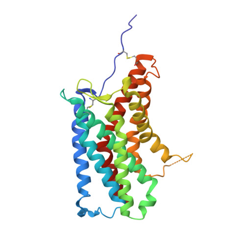
    The Duffy antigen receptor is a seven-transmembrane (7TM) protein expressed primarily at the surface of red blood cells and displays strikingly promiscuous binding to multiple inflammatory and homeostatic chemokines. It serves as the basis of the Duffy blood group system in humans and also acts as the primary attachment site for malarial parasite Plasmodium vivax and pore-forming toxins secreted by Staphylococcus aureus. Here, we comprehensively profile transducer coupling of this receptor, discover potential non-canonical signaling pathways, and determine the cryoelectron microscopy (cryo-EM) structure in complex with the chemokine CCL7. The structure reveals a distinct binding mode of chemokines, as reflected by relatively superficial binding and a partially formed orthosteric binding pocket. We also observe a dramatic shortening of TM5 and 6 on the intracellular side, which precludes the formation of the docking site for canonical signal transducers, thereby providing a possible explanation for the distinct pharmacological and functional phenotype of this receptor.


  • Organizational Affiliation

    Department of Biological Sciences and Bioengineering, Indian Institute of Technology Kanpur, Kanpur 208016, India.


Macromolecules
Find similar proteins by:  (by identity cutoff)  |  3D Structure
Entity ID: 1
MoleculeChains Sequence LengthOrganismDetailsImage
Atypical chemokine receptor 1A [auth B],
C [auth A]
267Homo sapiensMutation(s): 0 
Gene Names: ACKR1
Membrane Entity: Yes 
UniProt & NIH Common Fund Data Resources
Find proteins for Q16570 (Homo sapiens)
Explore Q16570 
Go to UniProtKB:  Q16570
PHAROS:  Q16570
GTEx:  ENSG00000213088 
Entity Groups  
Sequence Clusters30% Identity50% Identity70% Identity90% Identity95% Identity100% Identity
UniProt GroupQ16570
Sequence Annotations
Expand
  • Reference Sequence
Find similar proteins by:  (by identity cutoff)  |  3D Structure
Entity ID: 2
MoleculeChains Sequence LengthOrganismDetailsImage
C-C motif chemokine 7B [auth D],
D [auth C]
63Homo sapiensMutation(s): 0 
Gene Names: CCL7MCP3SCYA6SCYA7
Membrane Entity: Yes 
UniProt & NIH Common Fund Data Resources
Find proteins for P80098 (Homo sapiens)
Explore P80098 
Go to UniProtKB:  P80098
PHAROS:  P80098
GTEx:  ENSG00000108688 
Entity Groups  
Sequence Clusters30% Identity50% Identity70% Identity90% Identity95% Identity100% Identity
UniProt GroupP80098
Sequence Annotations
Expand
  • Reference Sequence
Experimental Data & Validation

Experimental Data

  • Method: ELECTRON MICROSCOPY
  • Resolution: 3.65 Å
  • Aggregation State: PARTICLE 
  • Reconstruction Method: SINGLE PARTICLE 
EM Software:
TaskSoftware PackageVersion
MODEL REFINEMENTPHENIX1.20.1
RECONSTRUCTIONcryoSPARC4.0

Structure Validation

View Full Validation Report


Entry History & Funding Information

Deposition Data


Funding OrganizationLocationGrant Number
Department of Biotechnology (DBT, India)IndiaBT/PR29041/BRB/10/1697/2018
Wellcome TrustUnited KingdomIA/S/20/1/504916
Science and Engineering Research Board (SERB)IndiaSPR/2020/000408
Science and Engineering Research Board (SERB)IndiaIPA/2020/000405

Revision History  (Full details and data files)

  • Version 1.0: 2024-07-31
    Type: Initial release
  • Version 1.1: 2024-08-14
    Changes: Data collection, Database references
  • Version 1.2: 2024-09-04
    Changes: Data collection, Database references