Structure and evolution of alanine/serine decarboxylases and the engineering of theanine production.
Wang, H., Zhu, B., Qiao, S., Dong, C., Wan, X., Gong, W., Zhang, Z.(2024) Elife 12
- PubMed: 39287621 Search on PubMedSearch on PubMed Central
- DOI: https://doi.org/10.7554/eLife.91046
- Primary Citation Related Structures:
8JG7, 8JIJ, 8JIK - PubMed Abstract:
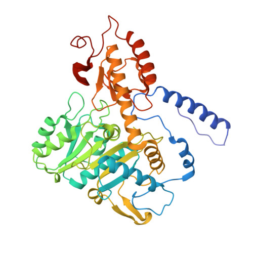
Ethylamine (EA), the precursor of theanine biosynthesis, is synthesized from alanine decarboxylation by alanine decarboxylase (AlaDC) in tea plants. AlaDC evolves from serine decarboxylase (SerDC) through neofunctionalization and has lower catalytic activity. However, lacking structure information hinders the understanding of the evolution of substrate specificity and catalytic activity. In this study, we solved the X-ray crystal structures of AlaDC from Camellia sinensis (CsAlaDC) and SerDC from Arabidopsis thaliana (AtSerDC). Tyr 341 of AtSerDC or the corresponding Tyr 336 of CsAlaDC is essential for their enzymatic activity. Tyr 111 of AtSerDC and the corresponding Phe 106 of CsAlaDC determine their substrate specificity. Both CsAlaDC and AtSerDC have a distinctive zinc finger and have not been identified in any other Group II PLP-dependent amino acid decarboxylases. Based on the structural comparisons, we conducted a mutation screen of CsAlaDC. The results indicated that the mutation of L110F or P114A in the CsAlaDC dimerization interface significantly improved the catalytic activity by 110% and 59%, respectively. Combining a double mutant of CsAlaDC L110F/P114A with theanine synthetase increased theanine production 672% in an in vitro system. This study provides the structural basis for the substrate selectivity and catalytic activity of CsAlaDC and AtSerDC and provides a route to more efficient biosynthesis of theanine.
- Department of Life Sciences and Medicine, University of Science and Technology of China, Hefei, China.
Organizational Affiliation: