8J7M | pdb_00008j7m

ion channel


Experimental Data Snapshot

  • Method: ELECTRON MICROSCOPY
  • Resolution: 3.10 Å
  • Aggregation State: PARTICLE 
  • Reconstruction Method: SINGLE PARTICLE 

wwPDB Validation   3D Report Full Report


This is version 1.1 of the entry. See complete history


Literature

Structural mechanism of voltage-gated sodium channel slow inactivation.

Chen, H.Xia, Z.Dong, J.Huang, B.Zhang, J.Zhou, F.Yan, R.Shi, Y.Gong, J.Jiang, J.Huang, Z.Jiang, D.

(2024) Nat Commun 15: 3691-3691

  • DOI: https://doi.org/10.1038/s41467-024-48125-3
  • Primary Citation of Related Structures:  
    8J7F, 8J7H, 8J7M

  • PubMed Abstract: 

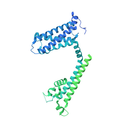
    Voltage-gated sodium (Na V ) channels mediate a plethora of electrical activities. Na V channels govern cellular excitability in response to depolarizing stimuli. Inactivation is an intrinsic property of Na V channels that regulates cellular excitability by controlling the channel availability. The fast inactivation, mediated by the Ile-Phe-Met (IFM) motif and the N-terminal helix (N-helix), has been well-characterized. However, the molecular mechanism underlying Na V channel slow inactivation remains elusive. Here, we demonstrate that the removal of the N-helix of Na V Eh (Na V Eh ΔN ) results in a slow-inactivated channel, and present cryo-EM structure of Na V Eh ΔN in a potential slow-inactivated state. The structure features a closed activation gate and a dilated selectivity filter (SF), indicating that the upper SF and the inner gate could serve as a gate for slow inactivation. In comparison to the Na V Eh structure, Na V Eh ΔN undergoes marked conformational shifts on the intracellular side. Together, our results provide important mechanistic insights into Na V channel slow inactivation.


  • Organizational Affiliation

    Department of Microbiology and Biotechnology, College of Life Sciences, Northeast Agricultural University, No. 600 Changjiang Road, Xiangfang District, Harbin, 150030, China.


Macromolecules
Find similar proteins by:  (by identity cutoff)  |  3D Structure
Entity ID: 1
MoleculeChains Sequence LengthOrganismDetailsImage
ion channel,Voltage dependent ion channel,Green fluorescent protein (Fragment),Voltage dependent ion channel,Green fluorescent protein (Fragment),Voltage dependent ion channel,Green fluorescent protein (Fragment)A,
B [auth D],
C [auth B],
D [auth C]
803Homo sapiensAequorea victoriaMutation(s): 0 
Membrane Entity: Yes 
UniProt
Find proteins for R1EKX3 (Emiliania huxleyi)
Explore R1EKX3 
Go to UniProtKB:  R1EKX3
Find proteins for R1FVI4 (Emiliania huxleyi)
Explore R1FVI4 
Go to UniProtKB:  R1FVI4
Entity Groups  
Sequence Clusters30% Identity50% Identity70% Identity90% Identity95% Identity100% Identity
UniProt GroupsR1EKX3R1FVI4
Sequence Annotations
Expand
  • Reference Sequence
Small Molecules
Ligands 2 Unique
IDChains Name / Formula / InChI Key2D Diagram3D Interactions
POV
Query on POV

Download Ideal Coordinates CCD File 
E [auth A],
H [auth D],
L [auth B],
O [auth C]
(2S)-3-(hexadecanoyloxy)-2-[(9Z)-octadec-9-enoyloxy]propyl 2-(trimethylammonio)ethyl phosphate
C42 H82 N O8 P
WTJKGGKOPKCXLL-PFDVCBLKSA-N
CLR
Query on CLR

Download Ideal Coordinates CCD File 
F [auth A]
G [auth A]
I [auth D]
J [auth D]
K [auth D]
CHOLESTEROL
C27 H46 O
HVYWMOMLDIMFJA-DPAQBDIFSA-N
Experimental Data & Validation

Experimental Data

  • Method: ELECTRON MICROSCOPY
  • Resolution: 3.10 Å
  • Aggregation State: PARTICLE 
  • Reconstruction Method: SINGLE PARTICLE 

Structure Validation

View Full Validation Report


Entry History & Funding Information

Deposition Data


Funding OrganizationLocationGrant Number
Brazilian National Council for Scientific and Technological Development (CNPq)Brazil31971134

Revision History  (Full details and data files)

  • Version 1.0: 2024-05-15
    Type: Initial release
  • Version 1.1: 2025-01-15
    Changes: Data collection, Database references, Structure summary