


Structure of CDK9/cyclin T1 in complex with inhibitor
Jiang, C., Ye, Y., Huang, Y.To be published.
Experimental Data Snapshot
Starting Model: experimental
View more details
Entity ID: 1 | |||||
|---|---|---|---|---|---|
| Molecule | Chains | Sequence Length | Organism | Details | Image |
| Cyclin-dependent kinase 9 | 332 | Homo sapiens | Mutation(s): 0 Gene Names: CDK9, CDC2L4, TAK EC: 2.7.11.22 (PDB Primary Data), 2.7.11.23 (PDB Primary Data) |  | |
UniProt & NIH Common Fund Data Resources | |||||
PHAROS: P50750 GTEx: ENSG00000136807 | |||||
Entity Groups | |||||
| Sequence Clusters | 30% Identity50% Identity70% Identity90% Identity95% Identity100% Identity | ||||
| UniProt Group | P50750 | ||||
Sequence AnnotationsExpand | |||||
Reference Sequence | |||||
Entity ID: 2 | |||||
|---|---|---|---|---|---|
| Molecule | Chains | Sequence Length | Organism | Details | Image |
| Cyclin-T1 | 259 | Homo sapiens | Mutation(s): 0 Gene Names: CCNT1 | 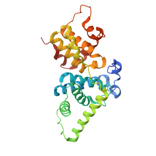 | |
UniProt & NIH Common Fund Data Resources | |||||
PHAROS: O60563 GTEx: ENSG00000129315 | |||||
Entity Groups | |||||
| Sequence Clusters | 30% Identity50% Identity70% Identity90% Identity95% Identity100% Identity | ||||
| UniProt Group | O60563 | ||||
Sequence AnnotationsExpand | |||||
Reference Sequence | |||||
| Ligands 1 Unique | |||||
|---|---|---|---|---|---|
| ID | Chains | Name / Formula / InChI Key | 2D Diagram | 3D Interactions | |
| NJ6 (Subject of Investigation/LOI) Download:Ideal Coordinates CCD File | C [auth A] | 2-[(4-azanylcyclohexyl)amino]-7-cyclopentyl-~{N},~{N}-dimethyl-pyrrolo[2,3-d]pyrimidine-6-carboxamide C20 H30 N6 O HZJPJDOVXYTUTI-SHTZXODSSA-N |  | ||
| Modified Residues 1 Unique | |||||
|---|---|---|---|---|---|
| ID | Chains | Type | Formula | 2D Diagram | Parent |
| TPO Query on TPO | A | L-PEPTIDE LINKING | C4 H10 N O6 P |  | THR |
| Length ( Å ) | Angle ( ˚ ) |
|---|---|
| a = 172.15 | α = 90 |
| b = 172.15 | β = 90 |
| c = 97.292 | γ = 120 |
| Software Name | Purpose |
|---|---|
| PHENIX | refinement |
| SCALA | data scaling |
| XDS | data reduction |
| PHENIX | phasing |
| Funding Organization | Location | Grant Number |
|---|---|---|
| Not funded | -- |


