8HHH

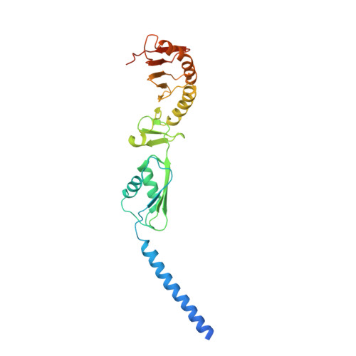
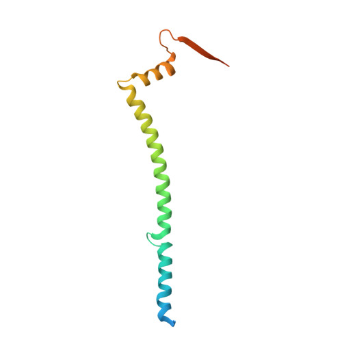
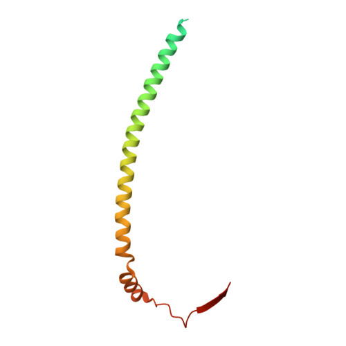
The bacterial divisome protein complex FtsB-FtsL-FtsQ


Experimental Data Snapshot

  • Method: X-RAY DIFFRACTION
  • Resolution: 3.30 Å
  • R-Value Free: 0.307 
  • R-Value Work: 0.276 
  • R-Value Observed: 0.277 

Starting Model: experimental
View more details

wwPDB Validation   3D Report Full Report


This is version 1.1 of the entry. See complete history


Literature

Structure of the heterotrimeric membrane protein complex FtsB-FtsL-FtsQ of the bacterial divisome.

Nguyen, H.T.V.Chen, X.Parada, C.Luo, A.C.Shih, O.Jeng, U.S.Huang, C.Y.Shih, Y.L.Ma, C.

(2023) Nat Commun 14: 1903-1903

  • DOI: https://doi.org/10.1038/s41467-023-37543-4
  • Primary Citation of Related Structures:  
    8HHF, 8HHG, 8HHH

  • PubMed Abstract: 

    The synthesis of the cell-wall peptidoglycan during bacterial cell division is mediated by a multiprotein machine, called the divisome. The essential membrane protein complex of FtsB, FtsL and FtsQ (FtsBLQ) is at the heart of the divisome assembly cascade in Escherichia coli. This complex regulates the transglycosylation and transpeptidation activities of the FtsW-FtsI complex and PBP1b via coordination with FtsN, the trigger for the onset of constriction. Yet the underlying mechanism of FtsBLQ-mediated regulation is largely unknown. Here, we report the full-length structure of the heterotrimeric FtsBLQ complex, which reveals a V-shaped architecture in a tilted orientation. Such a conformation could be strengthened by the transmembrane and the coiled-coil domains of the FtsBL heterodimer, as well as an extended β-sheet of the C-terminal interaction site involving all three proteins. This trimeric structure may also facilitate interactions with other divisome proteins in an allosteric manner. These results lead us to propose a structure-based model that delineates the mechanism of the regulation of peptidoglycan synthases by the FtsBLQ complex.


  • Organizational Affiliation

    Genomics Research Center, Academia Sinica, Taipei, 115, Taiwan.


Macromolecules
Find similar proteins by:  (by identity cutoff)  |  3D Structure
Entity ID: 1
MoleculeChains Sequence LengthOrganismDetailsImage
Cell division protein FtsQA [auth Q]276Escherichia coli K-12Mutation(s): 0 
Gene Names: 
Membrane Entity: Yes 
UniProt
Find proteins for P06136 (Escherichia coli (strain K12))
Explore P06136 
Go to UniProtKB:  P06136
Entity Groups  
Sequence Clusters30% Identity50% Identity70% Identity90% Identity95% Identity100% Identity
UniProt GroupP06136
Sequence Annotations
Expand
  • Reference Sequence
Find similar proteins by:  (by identity cutoff)  |  3D Structure
Entity ID: 2
MoleculeChains Sequence LengthOrganismDetailsImage
Cell division protein FtsB119Escherichia coli K-12Mutation(s): 1 
Gene Names: ygbQftsB
Membrane Entity: Yes 
UniProt
Find proteins for P0A6S5 (Escherichia coli (strain K12))
Explore P0A6S5 
Go to UniProtKB:  P0A6S5
Entity Groups  
Sequence Clusters30% Identity50% Identity70% Identity90% Identity95% Identity100% Identity
UniProt GroupP0A6S5
Sequence Annotations
Expand
  • Reference Sequence
Find similar proteins by:  (by identity cutoff)  |  3D Structure
Entity ID: 3
MoleculeChains Sequence LengthOrganismDetailsImage
Cell division protein FtsLC [auth L]121Escherichia coli K-12Mutation(s): 0 
Gene Names: ftsL
Membrane Entity: Yes 
UniProt
Find proteins for P0AEN4 (Escherichia coli (strain K12))
Explore P0AEN4 
Go to UniProtKB:  P0AEN4
Entity Groups  
Sequence Clusters30% Identity50% Identity70% Identity90% Identity95% Identity100% Identity
UniProt GroupP0AEN4
Sequence Annotations
Expand
  • Reference Sequence
Experimental Data & Validation

Experimental Data

  • Method: X-RAY DIFFRACTION
  • Resolution: 3.30 Å
  • R-Value Free: 0.307 
  • R-Value Work: 0.276 
  • R-Value Observed: 0.277 
  • Space Group: I 1 2 1
Unit Cell:
Length ( Å )Angle ( ˚ )
a = 100.29α = 90
b = 54.32β = 94.51
c = 179.261γ = 90
Software Package:
Software NamePurpose
PHENIXrefinement
XSCALEdata scaling
PDB_EXTRACTdata extraction
iMOSFLMdata reduction
PHENIXphasing

Structure Validation

View Full Validation Report


Entry History & Funding Information

Deposition Data


Funding OrganizationLocationGrant Number
Academia Sinica (Taiwan)Taiwan--

Revision History  (Full details and data files)

  • Version 1.0: 2023-04-26
    Type: Initial release
  • Version 1.1: 2023-10-18
    Changes: Data collection, Source and taxonomy