8GTD

Cryo-EM model of the marine siphophage vB_DshS-R4C portal-adaptor complex


Experimental Data Snapshot

  • Method: ELECTRON MICROSCOPY
  • Resolution: 4.70 Å
  • Aggregation State: PARTICLE 
  • Reconstruction Method: SINGLE PARTICLE 

wwPDB Validation   3D Report Full Report


This is version 1.1 of the entry. See complete history


Literature

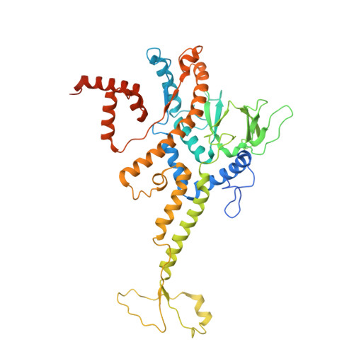
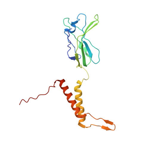
Structure and proposed DNA delivery mechanism of a marine roseophage.

Huang, Y.Sun, H.Wei, S.Cai, L.Liu, L.Jiang, Y.Xin, J.Chen, Z.Que, Y.Kong, Z.Li, T.Yu, H.Zhang, J.Gu, Y.Zheng, Q.Li, S.Zhang, R.Xia, N.

(2023) Nat Commun 14: 3609-3609

  • DOI: https://doi.org/10.1038/s41467-023-39220-y
  • Primary Citation of Related Structures:  
    8GTA, 8GTB, 8GTC, 8GTD, 8GTF

  • PubMed Abstract: 

    Tailed bacteriophages (order, Caudovirales) account for the majority of all phages. However, the long flexible tail of siphophages hinders comprehensive investigation of the mechanism of viral gene delivery. Here, we report the atomic capsid and in-situ structures of the tail machine of the marine siphophage, vB_DshS-R4C (R4C), which infects Roseobacter. The R4C virion, comprising 12 distinct structural protein components, has a unique five-fold vertex of the icosahedral capsid that allows genome delivery. The specific position and interaction pattern of the tail tube proteins determine the atypical long rigid tail of R4C, and further provide negative charge distribution within the tail tube. A ratchet mechanism assists in DNA transmission, which is initiated by an absorption device that structurally resembles the phage-like particle, RcGTA. Overall, these results provide in-depth knowledge into the intact structure and underlining DNA delivery mechanism for the ecologically important siphophages.


  • Organizational Affiliation

    State Key Laboratory of Molecular Vaccinology and Molecular Diagnostics, School of Public Health, School of Life Sciences, Xiamen University, Xiamen, 361102, China.


Macromolecules
Find similar proteins by:  (by identity cutoff)  |  3D Structure
Entity ID: 1
MoleculeChains Sequence LengthOrganismDetailsImage
Portal protein551Dinoroseobacter phage vB_DshS-R4CMutation(s): 0 
UniProt
Find proteins for A0A4Y6EI29 (Dinoroseobacter phage vB_DshS-R4C)
Explore A0A4Y6EI29 
Go to UniProtKB:  A0A4Y6EI29
Entity Groups  
Sequence Clusters30% Identity50% Identity70% Identity90% Identity95% Identity100% Identity
UniProt GroupA0A4Y6EI29
Sequence Annotations
Expand
  • Reference Sequence
Find similar proteins by:  (by identity cutoff)  |  3D Structure
Entity ID: 2
MoleculeChains Sequence LengthOrganismDetailsImage
Head-to-tail joining protein178Dinoroseobacter phage vB_DshS-R4CMutation(s): 0 
UniProt
Find proteins for A0A4Y6E755 (Dinoroseobacter phage vB_DshS-R4C)
Explore A0A4Y6E755 
Go to UniProtKB:  A0A4Y6E755
Entity Groups  
Sequence Clusters30% Identity50% Identity70% Identity90% Identity95% Identity100% Identity
UniProt GroupA0A4Y6E755
Sequence Annotations
Expand
  • Reference Sequence
Experimental Data & Validation

Experimental Data

  • Method: ELECTRON MICROSCOPY
  • Resolution: 4.70 Å
  • Aggregation State: PARTICLE 
  • Reconstruction Method: SINGLE PARTICLE 

Structure Validation

View Full Validation Report


Entry History & Funding Information

Deposition Data


Funding OrganizationLocationGrant Number
Not funded--

Revision History  (Full details and data files)

  • Version 1.0: 2023-07-12
    Type: Initial release
  • Version 1.1: 2024-06-19
    Changes: Data collection