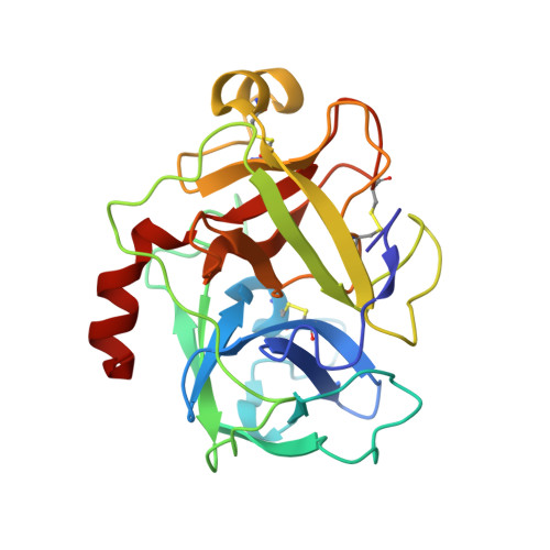
8G1V

Crystal Structure Matriptase (C731S) in Complex with Inhibitor MM1132-2


Experimental Data Snapshot

  • Method: X-RAY DIFFRACTION
  • Resolution: 1.35 Å
  • R-Value Free: 0.152 
  • R-Value Work: 0.131 
  • R-Value Observed: 0.132 

Starting Model: experimental
View more details

wwPDB Validation   3D Report Full Report


Ligand Structure Quality Assessment 


This is version 1.2 of the entry. See complete history


Literature

Use of protease substrate specificity screening in the rational design of selective protease inhibitors with unnatural amino acids: Application to HGFA, matriptase, and hepsin.

Mahoney, M.W.Helander, J.Kooner, A.S.Norman, M.Damalanka, V.C.De Bona, P.Kasperkiewicz, P.Rut, W.Poreba, M.Kashipathy, M.M.Battaile, K.P.Lovell, S.O'Donoghue, A.J.Craik, C.S.Drag, M.Janetka, J.W.

(2024) Protein Sci 33: e5110-e5110

  • DOI: https://doi.org/10.1002/pro.5110
  • Primary Citation of Related Structures:  
    8G1V

  • PubMed Abstract: 

    Inhibition of the proteolytic processing of hepatocyte growth factor (HGF) and macrophage stimulating protein (MSP) is an attractive approach for the drug discovery of novel anticancer therapeutics which prevent tumor progression and metastasis. Here, we utilized an improved and expanded version of positional scanning of substrate combinatorial libraries (PS-SCL) technique called HyCoSuL to optimize peptidomimetic inhibitors of the HGF/MSP activating serine proteases, HGFA, matriptase, and hepsin. These inhibitors have an electrophilic ketone serine trapping warhead and thus form a reversible covalent bond to the protease. We demonstrate that by varying the P2, P3, and P4 positions of the inhibitor with unnatural amino acids based on the protease substrate preferences learned from HyCoSuL, we can predictably modify the potency and selectivity of the inhibitor. We identified the tetrapeptide JH-1144 (8) as a single digit nM inhibitor of HGFA, matriptase and hepsin with excellent selectivity over Factor Xa and thrombin. These unnatural peptides have increased metabolic stability relative to natural peptides of similar structure. The tripeptide inhibitor PK-1-89 (2) has excellent pharmacokinetics in mice with good compound exposure out to 24 h. In addition, we obtained an X-ray structure of the inhibitor MM1132 (15) bound to matriptase revealing an interesting binding conformation useful for future inhibitor design.


  • Organizational Affiliation

    Department of Biochemistry and Molecular Biophysics, Washington University School of Medicine, Saint Louis, Missouri, USA.


Macromolecules
Find similar proteins by:  (by identity cutoff)  |  3D Structure
Entity ID: 1
MoleculeChains Sequence LengthOrganismDetailsImage
Suppressor of tumorigenicity 14 protein241Homo sapiensMutation(s): 1 
Gene Names: ST14PRSS14SNC19TADG15
EC: 3.4.21.109
UniProt & NIH Common Fund Data Resources
Find proteins for Q9Y5Y6 (Homo sapiens)
Explore Q9Y5Y6 
Go to UniProtKB:  Q9Y5Y6
PHAROS:  Q9Y5Y6
GTEx:  ENSG00000149418 
Entity Groups  
Sequence Clusters30% Identity50% Identity70% Identity90% Identity95% Identity100% Identity
UniProt GroupQ9Y5Y6
Sequence Annotations
Expand
  • Reference Sequence
Small Molecules
Ligands 4 Unique
IDChains Name / Formula / InChI Key2D Diagram3D Interactions
YIL (Subject of Investigation/LOI)
Query on YIL

Download Ideal Coordinates CCD File 
M [auth A]N~2~-{[3-(acetamidomethyl)phenyl]acetyl}-N-[(2S)-1-(1,3-benzothiazol-2-yl)-5-carbamimidamido-1,1-dihydroxypentan-2-yl]-L-leucinamide
C30 H41 N7 O5 S
PJRWAYIGCZNXHI-ZCYQVOJMSA-N
SO4
Query on SO4

Download Ideal Coordinates CCD File 
B [auth A],
C [auth A],
D [auth A],
E [auth A]
SULFATE ION
O4 S
QAOWNCQODCNURD-UHFFFAOYSA-L
GOL
Query on GOL

Download Ideal Coordinates CCD File 
K [auth A],
L [auth A]
GLYCEROL
C3 H8 O3
PEDCQBHIVMGVHV-UHFFFAOYSA-N
CL
Query on CL

Download Ideal Coordinates CCD File 
F [auth A],
G [auth A],
H [auth A],
I [auth A],
J [auth A]
CHLORIDE ION
Cl
VEXZGXHMUGYJMC-UHFFFAOYSA-M
Experimental Data & Validation

Experimental Data

  • Method: X-RAY DIFFRACTION
  • Resolution: 1.35 Å
  • R-Value Free: 0.152 
  • R-Value Work: 0.131 
  • R-Value Observed: 0.132 
  • Space Group: P 62 2 2
Unit Cell:
Length ( Å )Angle ( ˚ )
a = 149.473α = 90
b = 149.473β = 90
c = 58.501γ = 120
Software Package:
Software NamePurpose
PHENIXrefinement
Aimlessdata scaling
XDSdata reduction
PHASERphasing

Structure Validation

View Full Validation Report


Ligand Structure Quality Assessment 


Entry History & Funding Information

Deposition Data


Funding OrganizationLocationGrant Number
National Institutes of Health/National Institute Of Allergy and Infectious Diseases (NIH/NIAID)United StatesHHSN272201700059C
National Institutes of Health/Office of the DirectorUnited StatesS10OD030394

Revision History  (Full details and data files)

  • Version 1.0: 2024-07-10
    Type: Initial release
  • Version 1.1: 2024-08-07
    Changes: Database references
  • Version 1.2: 2024-08-14
    Changes: Database references