Use of protease substrate specificity screening in the rational design of selective protease inhibitors with unnatural amino acids: Application to HGFA, matriptase, and hepsin.
Mahoney, M.W., Helander, J., Kooner, A.S., Norman, M., Damalanka, V.C., De Bona, P., Kasperkiewicz, P., Rut, W., Poreba, M., Kashipathy, M.M., Battaile, K.P., Lovell, S., O'Donoghue, A.J., Craik, C.S., Drag, M., Janetka, J.W.(2024) Protein Sci 33: e5110-e5110
- PubMed: 39073183 Search on PubMedSearch on PubMed Central
- DOI: https://doi.org/10.1002/pro.5110
- Primary Citation Related Structures:
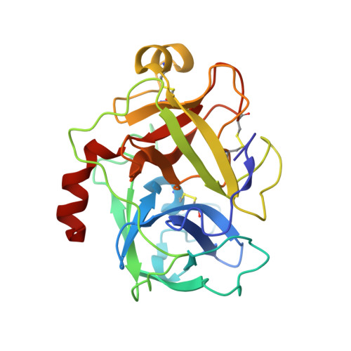
8G1V - PubMed Abstract:
Inhibition of the proteolytic processing of hepatocyte growth factor (HGF) and macrophage stimulating protein (MSP) is an attractive approach for the drug discovery of novel anticancer therapeutics which prevent tumor progression and metastasis. Here, we utilized an improved and expanded version of positional scanning of substrate combinatorial libraries (PS-SCL) technique called HyCoSuL to optimize peptidomimetic inhibitors of the HGF/MSP activating serine proteases, HGFA, matriptase, and hepsin. These inhibitors have an electrophilic ketone serine trapping warhead and thus form a reversible covalent bond to the protease. We demonstrate that by varying the P2, P3, and P4 positions of the inhibitor with unnatural amino acids based on the protease substrate preferences learned from HyCoSuL, we can predictably modify the potency and selectivity of the inhibitor. We identified the tetrapeptide JH-1144 (8) as a single digit nM inhibitor of HGFA, matriptase and hepsin with excellent selectivity over Factor Xa and thrombin. These unnatural peptides have increased metabolic stability relative to natural peptides of similar structure. The tripeptide inhibitor PK-1-89 (2) has excellent pharmacokinetics in mice with good compound exposure out to 24 h. In addition, we obtained an X-ray structure of the inhibitor MM1132 (15) bound to matriptase revealing an interesting binding conformation useful for future inhibitor design.
- Department of Biochemistry and Molecular Biophysics, Washington University School of Medicine, Saint Louis, Missouri, USA.
Organizational Affiliation: