Computational engineering of previously crystallized pyruvate formate-lyase activating enzyme reveals insights into SAM binding and reductive cleavage.
Moody, J.D., Hill, S., Lundahl, M.N., Saxton, A.J., Galambas, A., Broderick, W.E., Lawrence, C.M., Broderick, J.B.(2023) J Biological Chem 299: 104791-104791
- PubMed: 37156396 Search on PubMedSearch on PubMed Central
- DOI: https://doi.org/10.1016/j.jbc.2023.104791
- Primary Citation Related Structures:
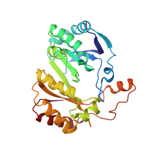
8FO0, 8FOL, 8FSI - PubMed Abstract:
Radical S-adenosyl-l-methionine (SAM) enzymes are ubiquitous in nature and carry out a broad variety of difficult chemical transformations initiated by hydrogen atom abstraction. Although numerous radical SAM (RS) enzymes have been structurally characterized, many prove recalcitrant to crystallization needed for atomic-level structure determination using X-ray crystallography, and even those that have been crystallized for an initial study can be difficult to recrystallize for further structural work. We present here a method for computationally engineering previously observed crystallographic contacts and employ it to obtain more reproducible crystallization of the RS enzyme pyruvate formate-lyase activating enzyme (PFL-AE). We show that the computationally engineered variant binds a typical RS [4Fe-4S] 2+/+ cluster that binds SAM, with electron paramagnetic resonance properties indistinguishable from the native PFL-AE. The variant also retains the typical PFL-AE catalytic activity, as evidenced by the characteristic glycyl radical electron paramagnetic resonance signal observed upon incubation of the PFL-AE variant with reducing agent, SAM, and PFL. The PFL-AE variant was also crystallized in the [4Fe-4S] 2+ state with SAM bound, providing a new high-resolution structure of the SAM complex in the absence of substrate. Finally, by incubating such a crystal in a solution of sodium dithionite, the reductive cleavage of SAM is triggered, providing us with a structure in which the SAM cleavage products 5'-deoxyadenosine and methionine are bound in the active site. We propose that the methods described herein may be useful in the structural characterization of other difficult-to-resolve proteins.
- Department of Chemistry and Biochemistry, Brigham Young University, Provo, Utah, USA; Department of Chemistry and Biochemistry, Montana State University, Bozeman, Montana, USA.
Organizational Affiliation: