8F42

Engineering Crystals with Tunable Symmetries from 14- or 16-Base-Long DNA Strands


Experimental Data Snapshot

  • Method: X-RAY DIFFRACTION
  • Resolution: 2.55 Å
  • R-Value Free: 0.279 
  • R-Value Work: 0.225 
  • R-Value Observed: 0.228 

Starting Model: in silico
View more details

wwPDB Validation   3D Report Full Report


Ligand Structure Quality Assessment 


This is version 1.2 of the entry. See complete history


Literature

Engineering DNA Crystals toward Studying DNA-Guest Molecule Interactions.

Zhang, C.Zhao, J.Lu, B.Seeman, N.C.Sha, R.Noinaj, N.Mao, C.

(2023) J Am Chem Soc 145: 4853-4859

  • DOI: https://doi.org/10.1021/jacs.3c00081
  • Primary Citation of Related Structures:  
    8EP8, 8EPB, 8EPD, 8EPE, 8EPF, 8EPG, 8EPI, 8F40, 8F42

  • PubMed Abstract: 

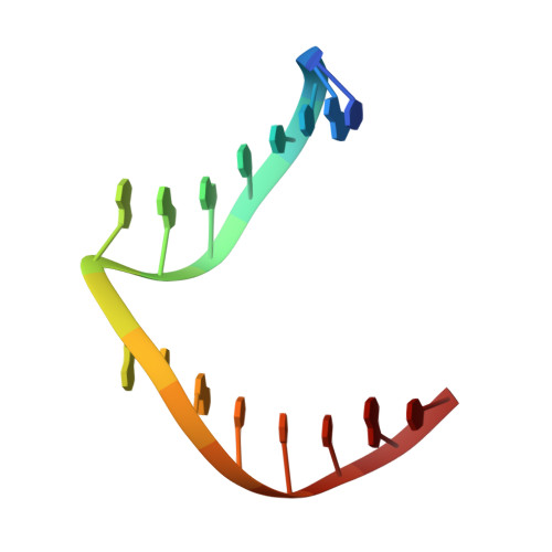
    Sequence-selective recognition of DNA duplexes is important for a wide range of applications including regulating gene expression, drug development, and genome editing. Many small molecules can bind DNA duplexes with sequence selectivity. It remains as a challenge how to reliably and conveniently obtain the detailed structural information on DNA-molecule interactions because such information is critically needed for understanding the underlying rules of DNA-molecule interactions. If those rules were understood, we could design molecules to recognize DNA duplexes with a sequence preference and intervene in related biological processes, such as disease treatment. Here, we have demonstrated that DNA crystal engineering is a potential solution. A molecule-binding DNA sequence is engineered to self-assemble into highly ordered DNA crystals. An X-ray crystallographic study of molecule-DNA cocrystals reveals the structural details on how the molecule interacts with the DNA duplex. In this approach, the DNA will serve two functions: (1) being part of the molecule to be studied and (2) forming the crystal lattice. It is conceivable that this method will be a general method for studying drug/peptide-DNA interactions. The resulting DNA crystals may also find use as separation matrices, as hosts for catalysts, and as media for material storage.


  • Organizational Affiliation

    Department of Chemistry, Purdue University, West Lafayette, Indiana 47907, United States.


Macromolecules

Find similar nucleic acids by:  Sequence   |   3D Structure  

Entity ID: 1
MoleculeChains LengthOrganismImage
DNA (5'-D(*CP*GP*CP*TP*TP*AP*AP*GP*GP*AP*AP*TP*TP*CP*GP*C)-3')
A, B
16synthetic construct
Sequence Annotations
Expand
  • Reference Sequence
Small Molecules
Ligands 1 Unique
IDChains Name / Formula / InChI Key2D Diagram3D Interactions
HT1 (Subject of Investigation/LOI)
Query on HT1

Download Ideal Coordinates CCD File 
C [auth B]2'-(4-ETHOXYPHENYL)-5-(4-METHYL-1-PIPERAZINYL)-2,5'-BI-BENZIMIDAZOLE
C27 H28 N6 O
PRDFBSVERLRRMY-UHFFFAOYSA-N
Experimental Data & Validation

Experimental Data

  • Method: X-RAY DIFFRACTION
  • Resolution: 2.55 Å
  • R-Value Free: 0.279 
  • R-Value Work: 0.225 
  • R-Value Observed: 0.228 
  • Space Group: P 43
Unit Cell:
Length ( Å )Angle ( ˚ )
a = 35.363α = 90
b = 35.363β = 90
c = 103.291γ = 90
Software Package:
Software NamePurpose
PHENIXrefinement
autoPROCdata reduction
STARANISOdata scaling
PHASERphasing

Structure Validation

View Full Validation Report


Ligand Structure Quality Assessment 


Entry History & Funding Information

Deposition Data


Funding OrganizationLocationGrant Number
National Science Foundation (NSF, United States)United StatesCMMI-2025187
National Science Foundation (NSF, United States)United StatesCCF-2107393
National Institutes of Health/National Institute of General Medical Sciences (NIH/NIGMS)United States1R01GM127884
National Institutes of Health/National Institute of General Medical Sciences (NIH/NIGMS)United States1R01GM127896

Revision History  (Full details and data files)

  • Version 1.0: 2023-03-08
    Type: Initial release
  • Version 1.1: 2023-03-15
    Changes: Database references
  • Version 1.2: 2024-04-03
    Changes: Data collection, Refinement description