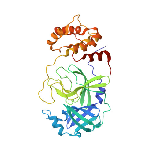
8E5C

Crystal Structure of SARS CoV-2 Mpro mutant L50F with Nirmatrelvir captured in two conformational states


Experimental Data Snapshot

  • Method: X-RAY DIFFRACTION
  • Resolution: 2.20 Å
  • R-Value Free: 0.254 
  • R-Value Work: 0.196 
  • R-Value Observed: 0.199 

wwPDB Validation   3D Report Full Report


Ligand Structure Quality Assessment 


This is version 1.2 of the entry. See complete history


Literature

Contributions of Hyperactive Mutations in M pro from SARS-CoV-2 to Drug Resistance.

Flynn, J.M.Zvornicanin, S.N.Tsepal, T.Shaqra, A.M.Kurt Yilmaz, N.Jia, W.Moquin, S.Dovala, D.Schiffer, C.A.Bolon, D.N.A.

(2024) ACS Infect Dis 10: 1174-1184

  • DOI: https://doi.org/10.1021/acsinfecdis.3c00560
  • Primary Citation of Related Structures:  
    8DT9, 8E4W, 8E5C

  • PubMed Abstract: 

    The appearance and spread of mutations that cause drug resistance in rapidly evolving diseases, including infections by the SARS-CoV-2 virus, are major concerns for human health. Many drugs target enzymes, and resistance-conferring mutations impact inhibitor binding or enzyme activity. Nirmatrelvir, the most widely used inhibitor currently used to treat SARS-CoV-2 infections, targets the main protease (M pro ) preventing it from processing the viral polyprotein into active subunits. Our previous work systematically analyzed resistance mutations in M pro that reduce binding to inhibitors; here, we investigate mutations that affect enzyme function. Hyperactive mutations that increase M pro activity can contribute to drug resistance but have not been thoroughly studied. To explore how hyperactive mutations contribute to resistance, we comprehensively assessed how all possible individual mutations in M pro affect enzyme function using a mutational scanning approach with a fluorescence resonance energy transfer (FRET)-based yeast readout. We identified hundreds of mutations that significantly increased the M pro activity. Hyperactive mutations occurred both proximal and distal to the active site, consistent with protein stability and/or dynamics impacting activity. Hyperactive mutations were observed 3 times more than mutations which reduced apparent binding to nirmatrelvir in recent studies of laboratory-grown viruses selected for drug resistance. Hyperactive mutations were also about three times more prevalent than nirmatrelvir binding mutations in sequenced isolates from circulating SARS-CoV-2. Our findings indicate that hyperactive mutations are likely to contribute to the natural evolution of drug resistance in M pro and provide a comprehensive list for future surveillance efforts.


  • Organizational Affiliation

    Department of Biochemistry and Molecular Biotechnology, University of Massachusetts Chan Medical School, Worcester, Massachusetts 01605, United States.


Macromolecules
Find similar proteins by:  (by identity cutoff)  |  3D Structure
Entity ID: 1
MoleculeChains Sequence LengthOrganismDetailsImage
3C-like proteinase nsp5306Severe acute respiratory syndrome coronavirus 2Mutation(s): 1 
Gene Names: rep1a-1b
EC: 3.4.22.69
UniProt
Find proteins for P0DTD1 (Severe acute respiratory syndrome coronavirus 2)
Explore P0DTD1 
Go to UniProtKB:  P0DTD1
Entity Groups  
Sequence Clusters30% Identity50% Identity70% Identity90% Identity95% Identity100% Identity
UniProt GroupP0DTD1
Sequence Annotations
Expand
  • Reference Sequence
Small Molecules
Ligands 4 Unique
IDChains Name / Formula / InChI Key2D Diagram3D Interactions
4WI (Subject of Investigation/LOI)
Query on 4WI

Download Ideal Coordinates CCD File 
C [auth A](1R,2S,5S)-N-{(1E,2S)-1-imino-3-[(3S)-2-oxopyrrolidin-3-yl]propan-2-yl}-6,6-dimethyl-3-[3-methyl-N-(trifluoroacetyl)-L-valyl]-3-azabicyclo[3.1.0]hexane-2-carboxamide
C23 H34 F3 N5 O4
WDVIRQQKRMIXGS-XIFHJVQQSA-N
DMS
Query on DMS

Download Ideal Coordinates CCD File 
B [auth A]DIMETHYL SULFOXIDE
C2 H6 O S
IAZDPXIOMUYVGZ-UHFFFAOYSA-N
CL
Query on CL

Download Ideal Coordinates CCD File 
J [auth A],
K [auth A],
L [auth A],
M [auth A]
CHLORIDE ION
Cl
VEXZGXHMUGYJMC-UHFFFAOYSA-M
NA
Query on NA

Download Ideal Coordinates CCD File 
D [auth A]
E [auth A]
F [auth A]
G [auth A]
H [auth A]
D [auth A],
E [auth A],
F [auth A],
G [auth A],
H [auth A],
I [auth A]
SODIUM ION
Na
FKNQFGJONOIPTF-UHFFFAOYSA-N
Experimental Data & Validation

Experimental Data

  • Method: X-RAY DIFFRACTION
  • Resolution: 2.20 Å
  • R-Value Free: 0.254 
  • R-Value Work: 0.196 
  • R-Value Observed: 0.199 
  • Space Group: P 21 21 2
Unit Cell:
Length ( Å )Angle ( ˚ )
a = 45.129α = 90
b = 63.693β = 90
c = 105.323γ = 90
Software Package:
Software NamePurpose
Cootmodel building
PHENIXrefinement
PHASERphasing
CrysalisProdata reduction
CrysalisProdata scaling
CrysalisProdata collection

Structure Validation

View Full Validation Report


Ligand Structure Quality Assessment 


Entry History & Funding Information

Deposition Data


Funding OrganizationLocationGrant Number
Other privateUnited States--

Revision History  (Full details and data files)

  • Version 1.0: 2024-02-07
    Type: Initial release
  • Version 1.1: 2024-03-27
    Changes: Database references
  • Version 1.2: 2024-04-24
    Changes: Database references