8DT9

Crystal Structure of SARS CoV-2 Mpro mutant L141R with Pfizer Intravenous Inhibitor PF-00835231


Experimental Data Snapshot

  • Method: X-RAY DIFFRACTION
  • Resolution: 2.00 Å
  • R-Value Free: 
    0.243 (Depositor), 0.240 (DCC) 
  • R-Value Work: 
    0.185 (Depositor), 0.180 (DCC) 
  • R-Value Observed: 
    0.188 (Depositor) 

wwPDB Validation   3D Report Full Report


Ligand Structure Quality Assessment 

Created with Raphaël 2.3.0Worse 01 BetterLigand structure goodness of fit to experimental dataBest fitted V2MClick on this verticalbar to view details

This is version 1.3 of the entry. See complete history


Literature

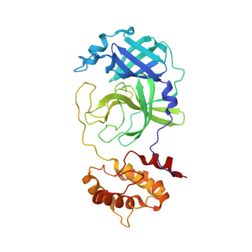
Contributions of Hyperactive Mutations in M pro from SARS-CoV-2 to Drug Resistance.

Flynn, J.M.Zvornicanin, S.N.Tsepal, T.Shaqra, A.M.Kurt Yilmaz, N.Jia, W.Moquin, S.Dovala, D.Schiffer, C.A.Bolon, D.N.A.

(2024) ACS Infect Dis 10: 1174-1184

  • DOI: https://doi.org/10.1021/acsinfecdis.3c00560
  • Primary Citation of Related Structures:  
    8DT9, 8E4W, 8E5C

  • PubMed Abstract: 

    The appearance and spread of mutations that cause drug resistance in rapidly evolving diseases, including infections by the SARS-CoV-2 virus, are major concerns for human health. Many drugs target enzymes, and resistance-conferring mutations impact inhibitor binding or enzyme activity. Nirmatrelvir, the most widely used inhibitor currently used to treat SARS-CoV-2 infections, targets the main protease (M pro ) preventing it from processing the viral polyprotein into active subunits. Our previous work systematically analyzed resistance mutations in M pro that reduce binding to inhibitors; here, we investigate mutations that affect enzyme function. Hyperactive mutations that increase M pro activity can contribute to drug resistance but have not been thoroughly studied. To explore how hyperactive mutations contribute to resistance, we comprehensively assessed how all possible individual mutations in M pro affect enzyme function using a mutational scanning approach with a fluorescence resonance energy transfer (FRET)-based yeast readout. We identified hundreds of mutations that significantly increased the M pro activity. Hyperactive mutations occurred both proximal and distal to the active site, consistent with protein stability and/or dynamics impacting activity. Hyperactive mutations were observed 3 times more than mutations which reduced apparent binding to nirmatrelvir in recent studies of laboratory-grown viruses selected for drug resistance. Hyperactive mutations were also about three times more prevalent than nirmatrelvir binding mutations in sequenced isolates from circulating SARS-CoV-2. Our findings indicate that hyperactive mutations are likely to contribute to the natural evolution of drug resistance in M pro and provide a comprehensive list for future surveillance efforts.


  • Organizational Affiliation

    Department of Biochemistry and Molecular Biotechnology, University of Massachusetts Chan Medical School, Worcester, Massachusetts 01605, United States.


Macromolecules
Find similar proteins by:  (by identity cutoff)  |  3D Structure
Entity ID: 1
MoleculeChains Sequence LengthOrganismDetailsImage
3C-like proteinase nsp5
A, B
306Severe acute respiratory syndrome coronavirus 2Mutation(s): 1 
Gene Names: rep1a-1b
EC: 3.4.22.69
UniProt
Find proteins for P0DTD1 (Severe acute respiratory syndrome coronavirus 2)
Explore P0DTD1 
Go to UniProtKB:  P0DTD1
Entity Groups  
Sequence Clusters30% Identity50% Identity70% Identity90% Identity95% Identity100% Identity
UniProt GroupP0DTD1
Sequence Annotations
Expand
  • Reference Sequence
Small Molecules
Ligands 3 Unique
IDChains Name / Formula / InChI Key2D Diagram3D Interactions
V2M (Subject of Investigation/LOI)
Query on V2M

Download Ideal Coordinates CCD File 
D [auth A],
H [auth B]
N-[(2S)-1-({(2S,3S)-3,4-dihydroxy-1-[(3S)-2-oxopyrrolidin-3-yl]butan-2-yl}amino)-4-methyl-1-oxopentan-2-yl]-4-methoxy-1H-indole-2-carboxamide
C24 H34 N4 O6
FDQSUXUTXIGUIA-PRIDNEQBSA-N
DMS
Query on DMS

Download Ideal Coordinates CCD File 
C [auth A],
G [auth B]
DIMETHYL SULFOXIDE
C2 H6 O S
IAZDPXIOMUYVGZ-UHFFFAOYSA-N
NA
Query on NA

Download Ideal Coordinates CCD File 
E [auth A]
F [auth A]
I [auth B]
J [auth B]
K [auth B]
SODIUM ION
Na
FKNQFGJONOIPTF-UHFFFAOYSA-N
Experimental Data & Validation

Experimental Data

  • Method: X-RAY DIFFRACTION
  • Resolution: 2.00 Å
  • R-Value Free:  0.243 (Depositor), 0.240 (DCC) 
  • R-Value Work:  0.185 (Depositor), 0.180 (DCC) 
  • R-Value Observed: 0.188 (Depositor) 
Space Group: P 1 21 1
Unit Cell:
Length ( Å )Angle ( ˚ )
a = 54.524α = 90
b = 98.467β = 107.452
c = 58.529γ = 90
Software Package:
Software NamePurpose
CrysalisProdata collection
PHENIXrefinement
CrysalisProdata collection
PHASERphasing
CrysalisProdata reduction
CrysalisProdata scaling
Cootmodel building

Structure Validation

View Full Validation Report


Ligand Structure Quality Assessment 

Created with Raphaël 2.3.0Worse 01 BetterLigand structure goodness of fit to experimental dataBest fitted V2MClick on this verticalbar to view details

Entry History & Funding Information

Deposition Data


Funding OrganizationLocationGrant Number
Other privateUnited States--

Revision History  (Full details and data files)

  • Version 1.0: 2024-02-07
    Type: Initial release
  • Version 1.1: 2024-03-27
    Changes: Database references
  • Version 1.2: 2024-04-24
    Changes: Database references
  • Version 1.3: 2024-10-23
    Changes: Structure summary