8DJD | pdb_00008djd

CRYSTAL STRUCTURE OF GLYCOGEN SYNTHASE KINASE 3 BETA COMPLEXED WITH 3-[(CYCLOPROPYLMETHYL)AMINO] -N-(4-PHENYLPYRIDIN-3-YL)IMIDAZO[1,2-B]PYRIDAZINE-8-CARBOX AMIDE


Experimental Data Snapshot

  • Method: X-RAY DIFFRACTION
  • Resolution: 2.21 Å
  • R-Value Free: 
    0.222 (Depositor), 0.213 (DCC) 
  • R-Value Work: 
    0.208 (Depositor) 
  • R-Value Observed: 
    0.209 (Depositor) 

Starting Model: other
View more details

wwPDB Validation 3D Report Full Report

Validation slider image for 8DJD

Ligand Structure Quality Assessment 


This is version 1.1 of the entry. See complete history

Literature

Structure-activity relationship (SAR) studies on substituted N-(pyridin-3-yl)-2-amino-isonicotinamides as highly potent and selective glycogen synthase kinase-3 (GSK-3) inhibitors.

Luo, G.Chen, L.Jacutin-Porte, S.Han, Y.Burton, C.R.Xiao, H.Krause, C.M.Cao, Y.Liu, N.Kish, K.Lewis, H.A.Macor, J.E.Dubowchik, G.M.

(2023) Bioorg Med Chem Lett 81: 129143-129143

  • DOI: https://doi.org/10.1016/j.bmcl.2023.129143
  • Primary Citation Related Structures: 
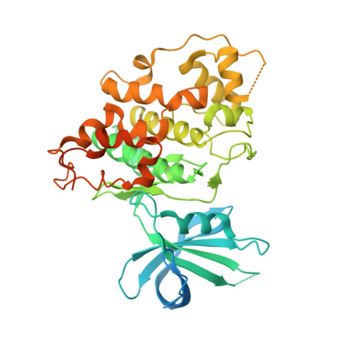
    8DJD

  • PubMed Abstract: 

    In our continuing efforts to explore structure-activity relationships around the novel class of potent, isonicotinamide-based GSK3 inhibitors described in our previous report, we extensively explored structural variations around both 4/5-pyridine substitutions and the amide group. Some analogs were found to have greatly improved pTau lowering potency while retaining high kinase selectivity. In contrast to previous active compounds 1a-c, a close analog 3h did not show in vivo efficacy in a triple-transgenic mouse Alzheimer's disease model. In general, these 2‑pyridinyl amide derivatives were prone to amidase mediated hydrolysis in mouse plasma.


  • Organizational Affiliation
    • Bristol-Myers Squibb, Wallingford, CT 06492, United States; Bristol Myers Squibb, Lawrenceville, NJ 08543, United States. Electronic address: guanglin.luo@bms.com.

Macromolecule Content 

  • Total Structure Weight: 99.31 kDa 
  • Atom Count: 5,500 
  • Modeled Residue Count: 679 
  • Deposited Residue Count: 882 
  • Unique protein chains: 1

Macromolecules

Find similar proteins by:|  3D Structure
Entity ID: 1
MoleculeChains  Sequence LengthOrganismDetailsImage
Glycogen synthase kinase-3 beta
A, B
441Homo sapiensMutation(s): 0 
Gene Names: GSK3B
EC: 2.7.11.26 (PDB Primary Data), 2.7.11.1 (PDB Primary Data)
UniProt & NIH Common Fund Data Resources
Find proteins for P49841 (Homo sapiens)
Explore P49841 
Go to UniProtKB:  P49841
PHAROS:  P49841
GTEx:  ENSG00000082701 
Entity Groups
Sequence Clusters30% Identity50% Identity70% Identity90% Identity95% Identity100% Identity
UniProt GroupP49841
Sequence Annotations
Expand
Reference Sequence

Small Molecules

Experimental Data & Validation

Experimental Data

  • Method: X-RAY DIFFRACTION
  • Resolution: 2.21 Å
  • R-Value Free:  0.222 (Depositor), 0.213 (DCC) 
  • R-Value Work:  0.208 (Depositor) 
  • R-Value Observed: 0.209 (Depositor) 
Space Group: P 21 21 21
Unit Cell:
Length ( Å )Angle ( ˚ )
a = 80.677α = 90
b = 83.659β = 90
c = 177.306γ = 90
Software Package:
Software NamePurpose
BUSTERrefinement
SCALAdata scaling
MOLREPphasing
PDB_EXTRACTdata extraction
XDSdata reduction

Structure Validation

View Full Validation Report


Ligand Structure Quality Assessment 


Entry History 

& Funding Information

Deposition Data


Funding OrganizationLocationGrant Number
Not fundedUnited States--

Revision History  (Full details and data files)

  • Version 1.0: 2023-03-22
    Type: Initial release
  • Version 1.1: 2024-04-03
    Changes: Data collection, Refinement description