8DFQ

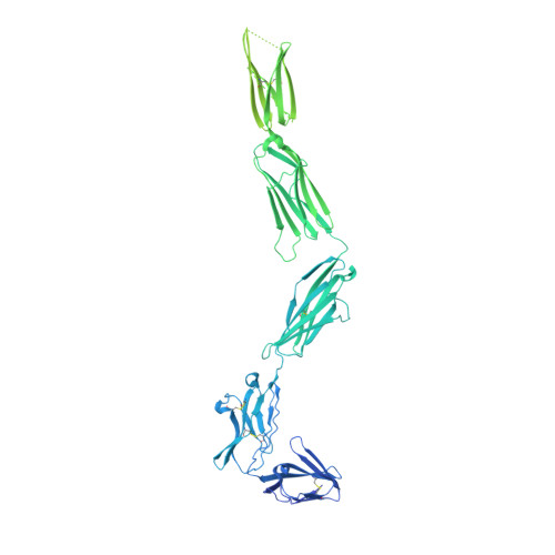
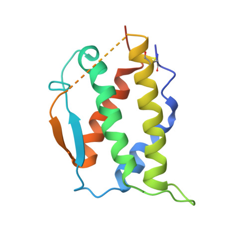
Ectodomain of full-length KIT(T417I,delta418-419)-SCF dimers


Experimental Data Snapshot

  • Method: ELECTRON MICROSCOPY
  • Resolution: 3.96 Å
  • Aggregation State: PARTICLE 
  • Reconstruction Method: SINGLE PARTICLE 

Starting Model: experimental
View more details

wwPDB Validation   3D Report Full Report


This is version 1.2 of the entry. See complete history


Literature

Cryo-EM analyses of KIT and oncogenic mutants reveal structural oncogenic plasticity and a target for therapeutic intervention.

Krimmer, S.G.Bertoletti, N.Suzuki, Y.Katic, L.Mohanty, J.Shu, S.Lee, S.Lax, I.Mi, W.Schlessinger, J.

(2023) Proc Natl Acad Sci U S A 120: e2300054120-e2300054120

  • DOI: https://doi.org/10.1073/pnas.2300054120
  • Primary Citation of Related Structures:  
    8DFM, 8DFP, 8DFQ

  • PubMed Abstract: 

    The receptor tyrosine kinase KIT and its ligand stem cell factor (SCF) are required for the development of hematopoietic stem cells, germ cells, and other cells. A variety of human cancers, such as acute myeloid leukemia, gastrointestinal stromal tumor, and mast cell leukemia, are driven by somatic gain-of-function KIT mutations. Here, we report cryo electron microscopy (cryo-EM) structural analyses of full-length wild-type and two oncogenic KIT mutants, which show that the overall symmetric arrangement of the extracellular domain of ligand-occupied KIT dimers contains asymmetric D5 homotypic contacts juxtaposing the plasma membrane. Mutational analysis of KIT reveals in D5 region an "Achilles heel" for therapeutic intervention. A ligand-sensitized oncogenic KIT mutant exhibits a more comprehensive and stable D5 asymmetric conformation. A constitutively active ligand-independent oncogenic KIT mutant adopts a V-shaped conformation solely held by D5-mediated contacts. Binding of SCF to this mutant fully restores the conformation of wild-type KIT dimers, including the formation of salt bridges responsible for D4 homotypic contacts and other hallmarks of SCF-induced KIT dimerization. These experiments reveal an unexpected structural plasticity of oncogenic KIT mutants and a therapeutic target in D5.


  • Organizational Affiliation

    Department of Pharmacology, Yale University School of Medicine, New Haven, CT 06520.


Macromolecules
Find similar proteins by:  (by identity cutoff)  |  3D Structure
Entity ID: 1
MoleculeChains Sequence LengthOrganismDetailsImage
Isoform 2 of Mast/stem cell growth factor receptor Kit
A, B
978Homo sapiensMutation(s): 2 
Gene Names: KITSCFR
EC: 2.7.10.1
UniProt & NIH Common Fund Data Resources
Find proteins for P10721 (Homo sapiens)
Explore P10721 
Go to UniProtKB:  P10721
PHAROS:  P10721
GTEx:  ENSG00000157404 
Entity Groups  
Sequence Clusters30% Identity50% Identity70% Identity90% Identity95% Identity100% Identity
UniProt GroupP10721
Glycosylation
Glycosylation Sites: 2Go to GlyGen: P10721-1
Sequence Annotations
Expand
  • Reference Sequence
Find similar proteins by:  (by identity cutoff)  |  3D Structure
Entity ID: 2
MoleculeChains Sequence LengthOrganismDetailsImage
Soluble KIT ligand
C, D
141Homo sapiensMutation(s): 0 
Gene Names: KITLGMGFSCF
UniProt & NIH Common Fund Data Resources
Find proteins for P21583 (Homo sapiens)
Explore P21583 
Go to UniProtKB:  P21583
PHAROS:  P21583
GTEx:  ENSG00000049130 
Entity Groups  
Sequence Clusters30% Identity50% Identity70% Identity90% Identity95% Identity100% Identity
UniProt GroupP21583
Sequence Annotations
Expand
  • Reference Sequence
Oligosaccharides

Help

Entity ID: 3
MoleculeChains Length2D Diagram Glycosylation3D Interactions
2-acetamido-2-deoxy-beta-D-glucopyranose-(1-4)-2-acetamido-2-deoxy-beta-D-glucopyranose
E, F, G
2N-Glycosylation
Glycosylation Resources
GlyTouCan:  G42666HT
GlyCosmos:  G42666HT
GlyGen:  G42666HT
Small Molecules
Ligands 1 Unique
IDChains Name / Formula / InChI Key2D Diagram3D Interactions
NAG
Query on NAG

Download Ideal Coordinates CCD File 
H [auth A]2-acetamido-2-deoxy-beta-D-glucopyranose
C8 H15 N O6
OVRNDRQMDRJTHS-FMDGEEDCSA-N
Experimental Data & Validation

Experimental Data

  • Method: ELECTRON MICROSCOPY
  • Resolution: 3.96 Å
  • Aggregation State: PARTICLE 
  • Reconstruction Method: SINGLE PARTICLE 
EM Software:
TaskSoftware PackageVersion
RECONSTRUCTIONcryoSPARC3.2.0
MODEL REFINEMENTPHENIX1.02.1-4487-000

Structure Validation

View Full Validation Report


Entry History & Funding Information

Deposition Data


Funding OrganizationLocationGrant Number
Not funded--

Revision History  (Full details and data files)

  • Version 1.0: 2023-03-29
    Type: Initial release
  • Version 1.1: 2023-04-05
    Changes: Database references
  • Version 1.2: 2024-10-30
    Changes: Data collection, Refinement description, Structure summary