8C59

CpG specific M.MpeI methyltransferase crystallized in the presence of 5-bromocytosine (converted to 5mC) and 5-methylcytosine containing dsDNA


Experimental Data Snapshot

  • Method: X-RAY DIFFRACTION
  • Resolution: 1.70 Å
  • R-Value Free: 0.193 
  • R-Value Work: 0.167 
  • R-Value Observed: 0.168 

Starting Model: experimental
View more details

wwPDB Validation   3D Report Full Report


Ligand Structure Quality Assessment 


This is version 1.1 of the entry. See complete history


Literature

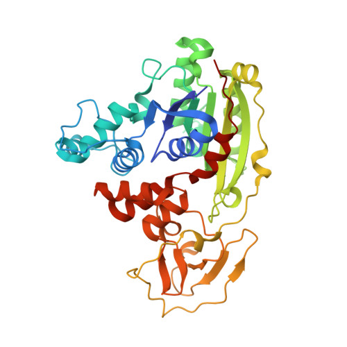
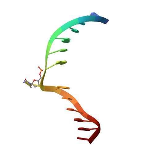
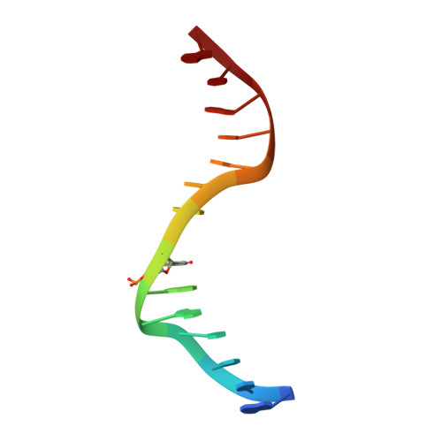
Cytosine analogues as DNA methyltransferase substrates.

Wojciechowski, M.Czapinska, H.Krwawicz, J.Rafalski, D.Bochtler, M.

(2024) Nucleic Acids Res 

  • DOI: https://doi.org/10.1093/nar/gkae568
  • Primary Citation of Related Structures:  
    8C56, 8C57, 8C58, 8C59

  • PubMed Abstract: 

    DNA methyltransferases are drug targets for myelodysplastic syndrome (MDS), chronic myelomonocytic leukemia (CMML), acute myelogenous leukemia (AML) and possibly β-hemoglobinopathies. We characterize the interaction of nucleoside analogues in DNA with a prokaryotic CpG-specific DNA methyltransferase (M.MpeI) as a model for mammalian DNMT1 methyltransferases. We tested DNA containing 5-hydroxymethylcytosine (5hmC), 5-hydroxycytosine (5OHC), 5-methyl-2-pyrimidinone (in the ribosylated form known as 5-methylzebularine, 5mZ), 5,6-dihydro-5-azacytosine (dhaC), 5-fluorocytosine (5FC), 5-chlorocytosine (5ClC), 5-bromocytosine (5BrC) and 5-iodocytosine (5IC). Covalent complex formation was by far most efficient for 5FC. Non-covalent complexes were most abundant for dhaC and 5mZ. Surprisingly, we observed methylation of 5IC and 5BrC, and to a lesser extent 5ClC and 5FC, in the presence, but not the absence of small molecule thiol nucleophiles. For 5IC and 5BrC, we demonstrated by mass spectrometry that the reactions were due to methyltransferase driven dehalogenation, followed by methylation. Crystal structures of M.MpeI-DNA complexes capture the 'in' conformation of the active site loop for analogues with small or rotatable (5mZ) 5-substituents and its 'out' form for bulky 5-substituents. Since very similar 'in' and 'out' loop conformations were also observed for DNMT1, it is likely that our conclusions generalize to other DNA methyltransferases.


  • Organizational Affiliation

    International Institute of Molecular and Cell Biology, Trojdena 4, 02-109 Warsaw, Poland.


Macromolecules

Find similar proteins by:  (by identity cutoff)  |  3D Structure
Entity ID: 1
MoleculeChains Sequence LengthOrganismDetailsImage
Cytosine-specific methyltransferase395Malacoplasma penetrans HF-2Mutation(s): 0 
Gene Names: MYPE4940
UniProt
Find proteins for Q8EVR5 (Malacoplasma penetrans (strain HF-2))
Explore Q8EVR5 
Go to UniProtKB:  Q8EVR5
Entity Groups  
Sequence Clusters30% Identity50% Identity70% Identity90% Identity95% Identity100% Identity
UniProt GroupQ8EVR5
Sequence Annotations
Expand
  • Reference Sequence

Find similar nucleic acids by:  Sequence   |   3D Structure  

Entity ID: 2
MoleculeChains LengthOrganismImage
DNA (5'-D(*CP*CP*AP*CP*AP*TP*GP*(5CM)P*GP*CP*TP*GP*AP*A)-3')14synthetic construct
Sequence Annotations
Expand
  • Reference Sequence

Find similar nucleic acids by:  Sequence   |   3D Structure  

Entity ID: 3
MoleculeChains LengthOrganismImage
DNA (5'-D(*GP*TP*TP*CP*AP*GP*(5CM)P*GP*CP*AP*TP*GP*TP*G)-3')14synthetic construct
Sequence Annotations
Expand
  • Reference Sequence
Small Molecules
Ligands 5 Unique
IDChains Name / Formula / InChI Key2D Diagram3D Interactions
SAM
Query on SAM

Download Ideal Coordinates CCD File 
D [auth A]S-ADENOSYLMETHIONINE
C15 H22 N6 O5 S
MEFKEPWMEQBLKI-FCKMPRQPSA-N
CIT
Query on CIT

Download Ideal Coordinates CCD File 
M [auth A]CITRIC ACID
C6 H8 O7
KRKNYBCHXYNGOX-UHFFFAOYSA-N
GOL
Query on GOL

Download Ideal Coordinates CCD File 
F [auth A]
G [auth A]
H [auth A]
I [auth A]
J [auth A]
F [auth A],
G [auth A],
H [auth A],
I [auth A],
J [auth A],
K [auth A],
L [auth A],
N [auth B],
O [auth C]
GLYCEROL
C3 H8 O3
PEDCQBHIVMGVHV-UHFFFAOYSA-N
CO3
Query on CO3

Download Ideal Coordinates CCD File 
P [auth C]CARBONATE ION
C O3
BVKZGUZCCUSVTD-UHFFFAOYSA-L
NA
Query on NA

Download Ideal Coordinates CCD File 
E [auth A]SODIUM ION
Na
FKNQFGJONOIPTF-UHFFFAOYSA-N
Experimental Data & Validation

Experimental Data

  • Method: X-RAY DIFFRACTION
  • Resolution: 1.70 Å
  • R-Value Free: 0.193 
  • R-Value Work: 0.167 
  • R-Value Observed: 0.168 
  • Space Group: P 41 21 2
Unit Cell:
Length ( Å )Angle ( ˚ )
a = 84.511α = 90
b = 84.511β = 90
c = 173.115γ = 90
Software Package:
Software NamePurpose
XDSdata reduction
XDSdata scaling
REFMACphasing
ARP/wARPmodel building
REFMACrefinement
PDB_EXTRACTdata extraction

Structure Validation

View Full Validation Report


Ligand Structure Quality Assessment 


Entry History & Funding Information

Deposition Data


Funding OrganizationLocationGrant Number
Foundation for Polish SciencePolandTEAM/2010-6/1
European Union (EU)European UnionTEAM/2010-6/1
Foundation for Polish SciencePolandPOIR.04.04.00-00-31DF/17-00
European Union (EU)European UnionPOIR.04.04.00-00-31DF/17-00
Polish National Science CentrePolandUMO-2014/13/B/NZ1/03991
Polish National Science CentrePolandUMO-2014/14/M/NZ5/00558
Polish National Science CentrePolandUMO-2018/30/Q/NZ2/00669
European Union (EU)European UnionPOIG.02.02.00-14-024/08-00

Revision History  (Full details and data files)

  • Version 1.0: 2024-01-17
    Type: Initial release
  • Version 1.1: 2024-07-17
    Changes: Database references