8BGT

O-Methyltransferase Plu4890 in complex with SAM


Experimental Data Snapshot

  • Method: X-RAY DIFFRACTION
  • Resolution: 2.15 Å
  • R-Value Free: 0.266 
  • R-Value Work: 0.223 
  • R-Value Observed: 0.225 

wwPDB Validation   3D Report Full Report


Ligand Structure Quality Assessment 


This is version 1.3 of the entry. See complete history


Literature

A set of closely related methyltransferases for site-specific tailoring of anthraquinone pigments.

Huber, E.M.Kreling, L.Heinrich, A.K.Dunnebacke, M.Pothig, A.Bode, H.B.Groll, M.

(2023) Structure 31: 573

  • DOI: https://doi.org/10.1016/j.str.2023.03.001
  • Primary Citation of Related Structures:  
    8BGT, 8BGX, 8BGY, 8BGZ, 8BH0, 8BIB, 8BIC, 8BID, 8BIE, 8BIF, 8BIG, 8BIH, 8BII, 8BIJ, 8BIR

  • PubMed Abstract: 

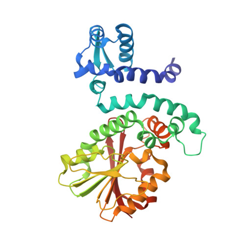
    Modification of the polyketide anthraquinone AQ-256 in the entomopathogenic Photorhabdus luminescens involves several O-methylations, but the biosynthetic gene cluster antA-I lacks corresponding tailoring enzymes. We here describe the identification of five putative, highly homologous O-methyltransferases encoded in the genome of P. luminescens. Activity assays in vitro and deletion experiments in vivo revealed that three of them account for anthraquinone tailoring by producing three monomethylated and two dimethylated species of AQ-256. X-ray structures of all five enzymes indicate high structural and mechanistic similarity. As confirmed by structure-based mutagenesis, a conserved histidine at the active site likely functions as a general base for substrate deprotonation and subsequent methyl transfer in all enzymes. Eight complex structures with AQ-256 as well as mono- and dimethylated derivatives confirm the substrate specificity patterns found in vitro and visualize how single amino acid differences in the active-site pockets impact substrate orientation and govern site-specific methylation.


  • Organizational Affiliation

    Technical University of Munich, TUM School of Natural Sciences, Department of Bioscience, Center for Protein Assemblies, Chair of Biochemistry, Ernst-Otto-Fischer-Str. 8, 85748 Garching, Germany. Electronic address: eva.huber@tum.de.


Macromolecules
Find similar proteins by:  (by identity cutoff)  |  3D Structure
Entity ID: 1
MoleculeChains Sequence LengthOrganismDetailsImage
Methyltransferase Plu4890
A, B
319Photorhabdus laumondii subsp. laumondii TTO1Mutation(s): 0 
UniProt
Find proteins for Q7MY08 (Photorhabdus laumondii subsp. laumondii (strain DSM 15139 / CIP 105565 / TT01))
Explore Q7MY08 
Go to UniProtKB:  Q7MY08
Entity Groups  
Sequence Clusters30% Identity50% Identity70% Identity90% Identity95% Identity100% Identity
UniProt GroupQ7MY08
Sequence Annotations
Expand
  • Reference Sequence
Small Molecules
Ligands 3 Unique
IDChains Name / Formula / InChI Key2D Diagram3D Interactions
SAM (Subject of Investigation/LOI)
Query on SAM

Download Ideal Coordinates CCD File 
C [auth A],
I [auth B]
S-ADENOSYLMETHIONINE
C15 H22 N6 O5 S
MEFKEPWMEQBLKI-FCKMPRQPSA-N
TRP
Query on TRP

Download Ideal Coordinates CCD File 
D [auth A]TRYPTOPHAN
C11 H12 N2 O2
QIVBCDIJIAJPQS-VIFPVBQESA-N
GOL
Query on GOL

Download Ideal Coordinates CCD File 
E [auth A],
F [auth A],
G [auth A],
H [auth A],
J [auth B]
GLYCEROL
C3 H8 O3
PEDCQBHIVMGVHV-UHFFFAOYSA-N
Experimental Data & Validation

Experimental Data

  • Method: X-RAY DIFFRACTION
  • Resolution: 2.15 Å
  • R-Value Free: 0.266 
  • R-Value Work: 0.223 
  • R-Value Observed: 0.225 
  • Space Group: C 1 2 1
Unit Cell:
Length ( Å )Angle ( ˚ )
a = 63.26α = 90
b = 76.78β = 96.67
c = 169.15γ = 90
Software Package:
Software NamePurpose
REFMACrefinement
PDB_EXTRACTdata extraction
XDSdata reduction
XSCALEdata scaling
CRANK2phasing

Structure Validation

View Full Validation Report


Ligand Structure Quality Assessment 


Entry History & Funding Information

Deposition Data


Funding OrganizationLocationGrant Number
Not funded--

Revision History  (Full details and data files)

  • Version 1.0: 2023-03-08
    Type: Initial release
  • Version 1.1: 2023-04-05
    Changes: Database references
  • Version 1.2: 2023-05-17
    Changes: Database references
  • Version 1.3: 2023-11-15
    Changes: Data collection, Source and taxonomy