8BFT

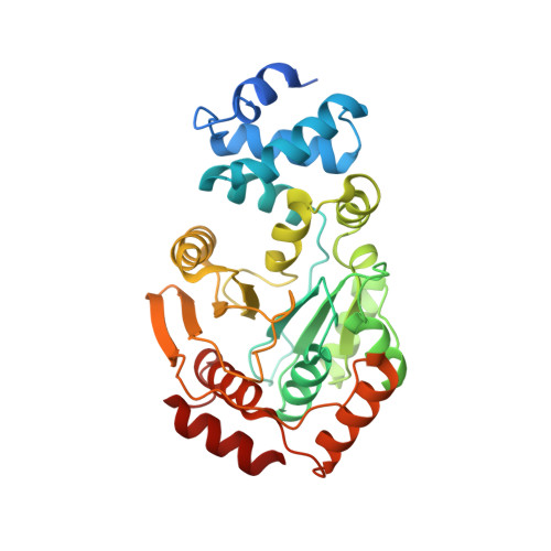
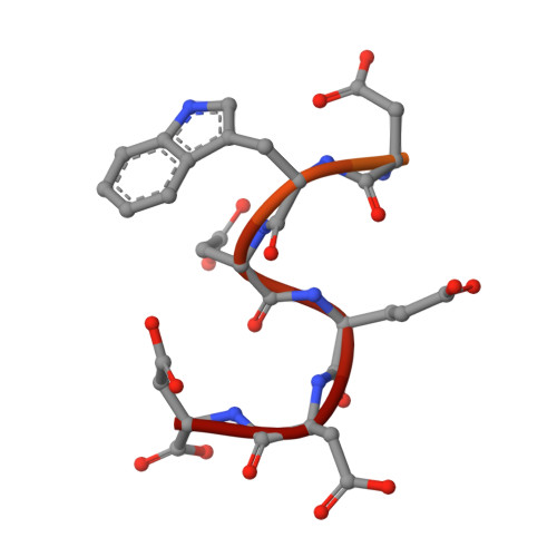
The E. coli TrpD2 protein YbiB in complex with a C-terminal peptide from ObgE


Experimental Data Snapshot

  • Method: X-RAY DIFFRACTION
  • Resolution: 1.19 Å
  • R-Value Free: 0.167 
  • R-Value Work: 0.137 
  • R-Value Observed: 0.138 

Starting Model: experimental
View more details

wwPDB Validation   3D Report Full Report


This is version 1.3 of the entry. See complete history


Literature

YbiB: a novel interactor of the GTPase ObgE.

Deckers, B.Vercauteren, S.De Kock, V.Martin, C.Lazar, T.Herpels, P.Dewachter, L.Verstraeten, N.Peeters, E.Ballet, S.Michiels, J.Galicia, C.Versees, W.

(2023) Nucleic Acids Res 51: 3420-3435

  • DOI: https://doi.org/10.1093/nar/gkad127
  • Primary Citation of Related Structures:  
    8BFR, 8BFT

  • PubMed Abstract: 

    Obg is a widely conserved and essential GTPase in bacteria, which plays a central role in a large range of important cellular processes, such as ribosome biogenesis, DNA replication, cell division and bacterial persistence. Nevertheless, the exact function of Obg in these processes and the interactions it makes within the associated pathways remain largely unknown. Here, we identify the DNA-binding TrpD2 protein YbiB as an interactor of the Escherichia coli Obg (ObgE). We show that both proteins interact with high affinity in a peculiar biphasic fashion, and pinpoint the intrinsically disordered and highly negatively charged C-terminal domain of ObgE as a main driver for this interaction. Molecular docking and X-ray crystallography, together with site-directed mutagenesis, are used to map the binding site of this ObgE C-terminal domain within a highly positively charged groove on the surface of the YbiB homodimer. Correspondingly, ObgE efficiently inhibits the binding of DNA to YbiB, indicating that ObgE competes with DNA for binding in the positive clefts of YbiB. This study thus forms an important step for the further elucidation of the interactome and cellular role of the essential bacterial protein Obg.


  • Organizational Affiliation

    VIB-VUB Center for Structural Biology, 1050 Brussels, Belgium.


Macromolecules
Find similar proteins by:  (by identity cutoff)  |  3D Structure
Entity ID: 1
MoleculeChains Sequence LengthOrganismDetailsImage
Protein YbiB343Escherichia coliMutation(s): 0 
Gene Names: ybiBb0800JW0785trpD2
UniProt
Find proteins for P30177 (Escherichia coli (strain K12))
Explore P30177 
Go to UniProtKB:  P30177
Entity Groups  
Sequence Clusters30% Identity50% Identity70% Identity90% Identity95% Identity100% Identity
UniProt GroupP30177
Sequence Annotations
Expand
  • Reference Sequence

Find similar proteins by:  Sequence   |   3D Structure  

Entity ID: 2
MoleculeChains Sequence LengthOrganismDetailsImage
GTPase ObgE/CgtA20Escherichia coliMutation(s): 0 
EC: 3.6.5
UniProt
Find proteins for P42641 (Escherichia coli (strain K12))
Explore P42641 
Go to UniProtKB:  P42641
Entity Groups  
Sequence Clusters30% Identity50% Identity70% Identity90% Identity95% Identity100% Identity
UniProt GroupP42641
Sequence Annotations
Expand
  • Reference Sequence
Experimental Data & Validation

Experimental Data

  • Method: X-RAY DIFFRACTION
  • Resolution: 1.19 Å
  • R-Value Free: 0.167 
  • R-Value Work: 0.137 
  • R-Value Observed: 0.138 
  • Space Group: C 1 2 1
Unit Cell:
Length ( Å )Angle ( ˚ )
a = 133.469α = 90
b = 63.895β = 97.35
c = 47.416γ = 90
Software Package:
Software NamePurpose
PHENIXrefinement
autoPROCdata reduction
autoPROCdata scaling
PHENIXphasing

Structure Validation

View Full Validation Report


Entry History & Funding Information

Deposition Data


Funding OrganizationLocationGrant Number
Research Foundation - Flanders (FWO)Belgium11D4621N
Vrije Universiteit Brussel (Blegium)BelgiumSRP50
Research Foundation - Flanders (FWO)BelgiumG0I1522N

Revision History  (Full details and data files)

  • Version 1.0: 2023-02-22
    Type: Initial release
  • Version 1.1: 2023-03-22
    Changes: Database references
  • Version 1.2: 2023-05-03
    Changes: Database references
  • Version 1.3: 2024-02-07
    Changes: Data collection